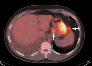
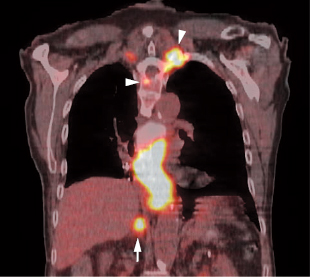
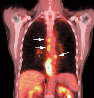
Gastric, Esophageal, and Gastrointestinal Stromal Tumors

17
Gastric, Esophageal, and
Gastrointestinal Stromal Tumors

Eugene C. Lin and Abass Alavi


Image Gastric Cancer


Clinical Indication: C


  1. Potential uses of positron emission tomography (PET) in patients with gastric cancer are staging, detecting recurrence, determining prognosis, and evaluating therapy response. However, the clinical role is not currently well defined.
  2. Prognosis

    1. Survival rate in patients with high fluorodeoxyglucose (FDG) uptake in the primary tumor is significantly lower than in patients with low FDG uptake.1 However, because mucinous and signet ring cell carcinomas typically have low FDG uptake, low FDG uptake does not neces sarily mean a better prognosis.
    2. A negative PET scan after surgical treatment with curative intent is associated with significantly longer survival.2

  3. Therapy response (early prediction). PET performed 14 days after initiation of chemotherapy predicts response to therapy.3

Accuracy and Comparison with Other Modalities


  1. PET. Sensitivity 71%, specificity 74 (advanced, metastatic, or recurrent gastric cancer)4
  2. Primary tumor. PET does not have a role in the detection of primary gastric cancer. Reported detection rates range from 17 (for tumor > 3 cm) to 96%.5,6

    1. A greater degree of FDG uptake is associated with greater depth of invasion, size of tumor, and lymph node metastases.1
    2. Signet ring cell and mucinous carcinomas have low FDG uptake.7,8

  3. Nodal metastases. The reported sensitivities of PET for detection of nodal metastases (Fig. 17.1) vary substantially (23 to 78%).1,4 The primary value of PET in detecting nodal metastases is its specificity (78 to 96%).4,5,9

    1. The accuracy of PET and computed tomography (CT) for detecting local and distant nodal metastases is similar.7
    2. CT is more sensitive in the detection of lymph node metastases in N1 and N2 disease, but PET is more specific.

  4. Body region7

    1. Accuracy for the primary lesion and for metastases to the liver (Fig. 17.2), lymph nodes, and lung is high.
    2. Accuracy for pleural and peritoneal disease and bone marrow metastases is low. Accuracy for small perigastric nodes can also be low if obscured by uptake in the primary tumor.

  5. Recurrent gastric cancer. Sensitivity 70%, specificity 69%3


image

Fig. 17.1 Gastric cancer. Axial positron emission tomography/computed tomography scan demonstrates uptake in a primary gastric cancer with two local nodal metastases (arrows).


Pearls and Pitfalls


  1. Histology. Signet ring cell and mucinous carcinomas have low FDG uptake.
  2. Morphology. The amount of uptake in the primary tumor may not correlate with histopathology.

    1. Poorly differentiated tumors may appear to have less uptake due to diffuse infiltration in the gastric wall.
    2. Well-differentiated tumors may appear to have more uptake due to mass formation.

  3. Other pathologies. Increased gastric FDG uptake can be secondary to etiologies other than gastric cancer.

    1. Diffuse increased uptake can be secondary to gastritis or lymphoma.
    2. Focal increased uptake can be secondary to gastric ulcer or lymphoma.

  4. Gastric remnant. Physiologic FDG uptake in a gastric remnant may be difficult to differentiate from recurrent tumor. Ingestion of water may be helpful. Gastric FDG uptake secondary to malignancy will persist after water ingestion.10

Image Detection of Primary Esophageal Cancer


Clinical Indication: D

PET has a limited role in evaluating primary esophageal tumors.


  1. Detection

    1. PET can detect primary tumors with depth of invasion of T1b or greater, but in situ and T1a tumors are not detectable.11
    2. The overall detection rate of PET for primary esophageal carcinoma is 80%. However, this depends on the T stage. The detection rate for T3 and T4 tumors is close to 100%, but the detection rate for T1 tumors is 43%.12
    3. PET cannot determine T stage.

  2. Level of uptake13

    1. The amount of uptake correlates positively with depth of tumor invasion, presence of lymph node metastases, and lymphatic invasion.
    2. Adenocarcinomas and squamous cell car cinomas overall have similar degrees of FDG uptake, although adenocarcinomas at or near the gastroesophageal junction often have lower uptake secondary to diffuse growth pattern and/or mucinous histopathology.14,15
    3. The majority of the evidence suggests that the survival rate with high FDG up take in the primary tumor is lower, but not all studies support this.14

Pitfalls


  1. Mild focal uptake at the gastroesophageal junction could be secondary to esophagitis, or it could be a normal variant (see Fig. 6.27, p. 56). An SUV > 4 at the gastroesophageal junction is concerning for malignancy.16
  2. Hiatal hernias can cause large areas of uptake at the gastroesophageal junction.
  3. Benign strictures can have substantial FDG uptake after dilatation.
  4. Esophageal leiomyomas can have FDG uptake.17
  5. Respiratory misregistration. Respiratory misregistration artifact on PET/CT (which is greatest in the peridiaphragmatic region) may result in erroneous SUV measurements of distal esophageal tumors.18


image

Fig. 17.4 Metastatic esophageal cancer. Coronal positron emission tomography/computed tomography scan in a patient with esophageal carcinoma demonstrates intense uptake in the primary tumor and adrenal (arrow) and bone (arrowheads) metastases.


image

Fig. 17.5 Esophageal cancer with locoregional nodal metastases. Coronal positron emission tomography/computed tomography (PET/CT) scan demonstrates multiple metastases to locoregional paraesophageal nodes (arrows) from a distal esophageal cancer. Although endoscopic ultrasound would usually be more sensitive for detection of these nodes, a positive PET study is more specific for metastatic disease.


Table 17.1 Sensitivity and Specificity of Positron Emission Tomography (PET) versus Computed Tomography (CT) and Endoscopic Ultrasound (EUS) in the Detection of Locoregional Nodal Disease in Esophageal Cancer

Sensitivity % Specificity %
PET 22 91
CT/EUS 83 45


Table 17.2 Sensitivity and Specificity of Positron Emission Tomography (PET) versus Computed Tomography (CT) and Endoscopic Ultrasound (EUS) in the Detection of Distant Nodes in Esophageal Cancer

Sensitivity % Specificity %
PET 77 90
CT/EUS 46 69

Image Staging of Esophageal Cancer


Clinical Indication: A


  1. The combination of PET and endoscopic ultrasound (EUS) can be the most cost-effective method of staging esophageal cancer.19
  2. The primary value of PET is in20

    1. Detecting distant metastases (Figs. 17.3 and 17.4)
    2. Improving the specificity of lymph node staging

  3. However, PET may not be routinely useful in patients with early-stage esophageal cancer (T ≥ 2), as these patients have a low incidence of lymphatic metastases.21
  4. The overall incremental benefit in staging accuracy of PET compared with CT is 14%.12 PET will identify unsuspected distant metastatic disease in 5 to 8%22 of patients without evidence of metastases after conventional work-up.
  5. Prognosis. Greater tumor length on PET and increased number of PET positive lymph nodes predict low survival rate.23


Table 17.3 Sensitivity and Specificity of Positron Emission Tomography (PET) versus Computed Tomography (CT) in the Detection of Distant Metastases (Nodal and Other) in Esophageal Cancer

Sensitivity % Specificity %
PET 69 93
CT 46 74


Table 17.4 Sensitivity and Specificity of Positron Emission Tomography (PET) versus Bone Scan in the Detection of Bone Metastases in Esophageal Cancer

Sensitivity % Specificity %
PET 92 94
Bone scan 77 84

Accuracy24


  1. PET: locoregional. Sensitivity 51%, specificity 94%
  2. PET: distant metastases. Sensitivity 67%, specificity 97%
  3. PET/CT: locoregional. Sensitivity 94%, specificity 92%25

    • Significantly more accurate than PET alone

Comparison with Other Modalities


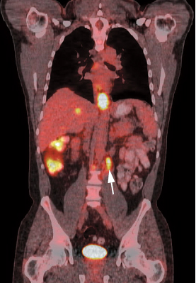
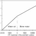
  1. Locoregional nodes.21 PET is insensitive for locoregional disease and cannot replace CT/EUS for locoregional staging, but a positive result is more specific than CT/EUS for nodal disease (Fig. 17.5). A large percentage of false-negative nodal groups on PET are in the immediate vicinity of the primary tumor.12 EUS will detect more pathologic periesophageal and celiac axis nodes than PET or CT (Table 17.1).26
  2. Distant nodes (Table 17.2).21
  3. Distant metastases (nodal and other) (Table 17.3)27
  4. Bone metastases. PET may be more accurate than bone scintigraphy for bone metas-tases (Table 17.4).28
  5. Anatomical region. PET has the highest accuracy in the neck, upper thoracic, and abdominal regions but low sensitivity in the mid- and lower thoracic regions.13


image

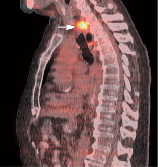
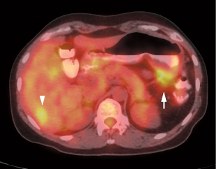
Fig. 17.6 Esophageal cancer recurrence. Sagittal positron emission tomography/computed tomography (PET/CT) in an esophageal cancer patient status postesophagectomy and gastric pull-through demonstrates recurrence at the proximal anastomosis (arrow).

Only gold members can continue reading. Log In or Register to continue

Stay updated, free articles. Join our Telegram channel

Sep 3, 2016 | Posted by in GENERAL RADIOLOGY | Comments Off on Gastric, Esophageal, and Gastrointestinal Stromal Tumors

Full access? Get Clinical Tree

Get Clinical Tree app for offline access