Fig. 22.1
Weight-lifting populations. Strength athletes can be categorized by their level of experience. The incidence of injuries is highest among beginner or inexperienced lifters with accidental trauma being most common. Elite competitors also have a higher incidence of injuries because these athletes strive for maximal performance which stresses the bones and muscles
In general, the majority of lifting injuries relate to either tensile forces or compressive forces, with overuse contributing a significant component to the injury. This is particularly true of elite athletes. While some weight-lifting injuries are readily apparent at the time of the injury, it is noteworthy that most injuries are difficult to isolate or quantify either clinically or with the use of radiographs. In these situations, MRI is best suited for further evaluation (Boutin et al. 2002; Bencardino et al. 2000). The tendon, myotendinous unit, muscle, and bones are characteristic target sites, and several specific injuries related to the sport of weight lifting will be covered in this chapter.
22.1.2 Types of Strength Athletes
There are five distinct groups of strength athletes, and though they may share overlapping areas of risk, it is important to recognize that each group may have a higher incidence for a specific injury common to that group’s style of lifting (Lavallee and Balam 2010). Olympic-style weight lifting is a sport that focuses on lifting a weight above the head either in a single-stage motion as in the snatch or in two-stage motion as in the clean and jerk, where the weight temporarily rests on the shoulders during its upward movement (Fig. 22.2). Power lifting involves three disciplines, the squat, bench press, and dead lift, which are basic motions that are performed by elite strength athletes as well as “weekend warriors” (Fig. 22.3). Strength specialists or style-dependent strength sports, people who compete in “strongman” competitions, subject their bodies to carrying, pushing, and lifting activities that are considered normal motions of life but which are taken to an extreme or maximal load. A growing number of people have been participating in cross fit competitions that incorporate endurance and strength maneuvers that require an athlete to balance aerobic with anaerobic conditioning. Bodybuilders are athletes who use weight lifting to enhance the muscularity in their physique. The last group are athletes who use resistance training as part of a core of exercises that is used to enhance overall athletic performance.
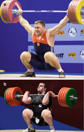
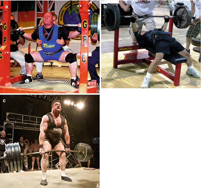
Fig. 22.2
Olympic weight lifting. (a) There are two disciplines in Olympic weight lifting. In the “snatch,” the lifter attempts to move the barbell above his head in one swift motion. (b) In the “clean and jerk,” there is an intervening step shown here where the barbell rests on the shoulders prior to it being lifted above the head
Fig. 22.3
Powerlifting weight lifting. (a) There are three lifts in powerlifting. In the “squat,” the lifter descends with a barbell behind the shoulders until the top of the thighs is parallel with the floor. (b) In the “bench press,” the barbell descends to the chest and then is pushed back up until the arms are fully extended, as shown by one of the authors during a competition. (c) In the “dead lift,” the barbell is lifted off the floor until the lifter achieves an upright position with the legs locked in full extension
22.2 Mechanisms of Injury
Injuries related to weight lifting can affect any tissue. Weight-lifting injuries that affect the musculoskeletal system are primarily categorized as injuries that involve the myotendinous unit, joints, or the osseous skeleton. Injuries affecting the ligaments are uncommon and usually are associated with dislocations.
22.2.1 Myotendinous Unit
A tear in the myotendinous unit occurs when tension in the unit exceeds the strength of its weakest structural element. The point of failure is dependent on several factors including preexisting conditions such as tendinosis, the position of overload, and the velocity of injury (Yu and Habib 2005; Sharafi et al. 2011). Myotendinous injuries constitute about two-thirds of all injuries and can affect both the highly trained athlete and the novice lifter.
The myotendinous unit is composed of muscle, the myotendinous junction, and tendon. The basic function of muscles is to produce joint motion by creating tensile forces that are transmitted through the myotendinous junction to the origin and insertion of tendons. There are two types of muscle contraction, eccentric and concentric contractions (Shellock et al. 1991). In eccentric contraction, the muscle fibers lengthen when the resisting force is greater than the force generated by the muscle (El-Khoury et al. 1996). Concentric contraction occurs when the resisting load is less than the force generated by the muscle, shortening the muscle. Eccentric muscle activation increases the potential for injury, particularly when it is acting to absorb kinetic energy, because it creates greater tensile forces within the muscle than can be produced during concentric contraction (Garrett 1990). Prolonged eccentric contraction increases the incidence of delayed-onset muscle soreness (DOMS) characterized by pain, acute inflammation, and cellular infiltration (Fig. 22.4). Muscles that extend across two joints are at most risk for strains because their primary function is eccentric contraction. Muscles that are particularly susceptible to injury include the biceps brachii, rectus femoris, biceps femoris, semimembranosus, semitendinosus, adductor, vastus medialis, soleus, and medial gastrocnemius muscles (Palmer et al. 1999).
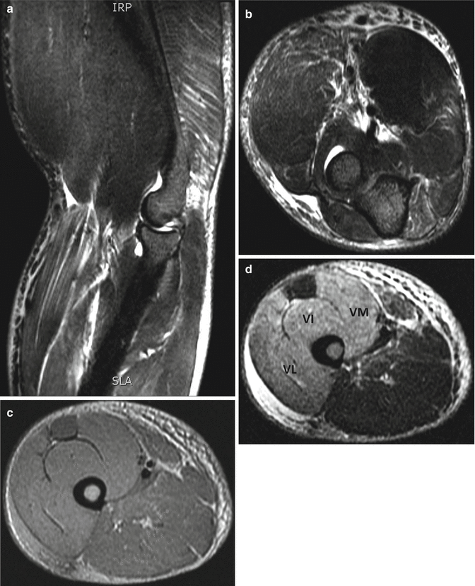
Fig. 22.4
Two patients with delayed-onset muscle soreness (DOMS). Sagittal (a) and axial (b) fat-suppressed T2-weighted MRI of the elbow show diffuse interstitial edema in the triceps, biceps, brachioradialis, and forearm musculature after working out arms to maximal exertion. The athlete had intense muscle pain for several days. Axial fat-suppressed proton density-weighted (c) and T2-weighted (d) MRI of the distal thigh show architectural distortion and marked edema in the majority of the quadriceps musculature as well as subcutaneous fat edema from performing multiple “negative” knee extensions or eccentric contraction against gravity. VL vastus lateralis, VI vastus intermedius, VM vastus medialis
The composition of a muscle can also increase its risk for injury. Differences in muscle fiber types reflect the speed of contraction and the endurance of the muscle (Garrett et al. 1984). A type 1 fiber, considered slow twitch, has slow contraction and relaxation times but is resistant to fatigue. A type 2 fiber, considered fast twitch, has fast contraction and relaxation times and is well suited for intense activities of short duration. Muscles with a high proportion of type 2 fibers are capable of generating more force but are also more susceptible to fatigue. Fatigued muscles absorb less energy and are more likely to sustain an injury.
The myotendinous junction is a specialized region of highly folded membranes at the muscle-tendon interface. This folding increases the junctional surface area by 10–20 times, which decreases the stress per unit area (El-Khoury et al. 1996). But because the capacity for energy absorption is less than either muscle or tendon, it is more susceptible to strains. A normal tendon does not rupture when subjected to extreme loading, but muscles subjected to forceful stretching consistently disrupt near the muscle-tendon junction or near the bone-tendon junction (Towers et al. 2003). For a tendon to fail in its mid-substance, an underlying abnormality typically already exists (Kannus and Natri 1997). Cyclical loading can promote fatigue-induced damage resulting in microstructural, biological, and mechanical changes to the tendon that correlates with histologic tendinopathy (Andarawis-Puri and Flatow 2011; Shepherd and Screen 2013; Andarawis-Puri et al. 2012).
MRI is the preferred imaging method for evaluating muscle and myotendinous injuries (Goodwin 2011; Lee et al. 2012). Because of its unparalleled anatomic resolution, it depicts a high sensitivity to acute and chronic muscle abnormalities (Shelly et al. 2009). Areas of intramuscular hemorrhage correlate well with histopathological changes in the muscle (Yan et al. 2013). Additionally, measurements related to the extent of injury such as cross-sectional area and longitudinal extent of muscle injury adjacent to the myotendinous junction have been shown to correlate with prognosis and recovery time needed for the athlete prior to return to competition (Slavotinek 2010; Blankenbaker and Tuite 2010).
22.2.2 Types of Strains
The myotendinous junction is the site of most strains although the muscle alone can also be injured especially in broad muscles. Pathologically, a strained muscle demonstrates a combination of torn fibers, inflammation, edema, and hemorrhage with protein degradation and regeneration. The severity of an acute strain depends on the rate, magnitude, and duration of stress loading (Steinbach et al. 1997; Fleckenstein et al. 1989). A strain of the muscle is graded according to loss of function and strength and the area of tissue involved. A first-degree strain of the myotendinous junction is considered a stretching injury without tearing and is characterized by edema and hemorrhage surrounding the myotendinous junction and edema tracking along the muscle fascicles and interstitial connective tissue (Deutsch and Mink 1989). This type of injury typically heals without residual damage, and the imaging findings tend to resolve completely. A first-degree strain of the muscle produces minimal disruption of the tissue and is associated with less than 5 % loss of function. A diffuse, infiltrative pattern of edema and hemorrhage characterizes the injury, appearing similar to a muscle contusion (Fig. 22.5).
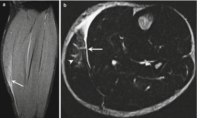
Fig. 22.5
Grade 1 strain of the gastrocnemius muscle. (a) Coronal STIR MRI shows loss of contour in the medial aspect of the medial gastrocnemius muscle (arrow). (b) Axial T2-weighted MRI shows focal, feathery increased signal intensity (arrowhead) consistent with edema/hemorrhage. There is also rupture of the anterior aponeurosis (arrow) with fluid in the interstitial tissues between the gastrocnemius and soleus muscles
A second-degree strain of the myotendinous junction represents a partial tear and is characterized by irregular thinning and laxity of the tendon owing to partial disruption of the fibers. Edema and hemorrhage are more pronounced, becoming interspersed within the fascial planes between the muscle fascicles and muscle groups (Fig. 22.6) (Fleckenstein et al. 1989; De Smet 1993). This injury is often associated with the formation of a hematoma which may lead to the formation of a peripheral rim of hemosiderin within one week from the time of injury (Lee et al. 2011). Restoration of function is largely dependent on the magnitude of fiber disruption, but conservative treatment is often sufficient to reestablish normal strength and range of motion. A second-degree strain of the muscle is associated with more pronounced loss of muscle strength which is dependent on the volume of fiber separation. Imaging findings remain abnormal for a much longer period of time.
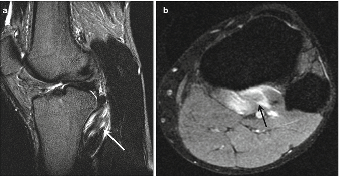
Fig. 22.6
Grade 2 strain of the popliteus muscle. Sagittal (a) and axial (b) fat-suppressed T2-weighted MRI of the knee shows localized high signal intensity in the popliteus muscle (arrows) indicating significant muscle fiber disruption
Third-degree strains that occur at the myotendinous junction represent a complete rupture of the tendon, enthesis or an osseous avulsion at the tendinous attachment (Steinbach et al. 1997). A complete rupture is often accompanied by tendon retraction becoming more pronounced with delayed diagnosis (Fig. 22.7). Edema may be sufficiently severe in the acute phase of the injury to obscure visualization of the anatomic structures, rendering it difficult to differentiate a complete tear from a large partial tear. Clinically, muscle paresis is evident. Restoration of function requires surgery. Otherwise, the muscle atrophies and becomes infiltrated with lipoid tissue (Kuzel et al. 2013).
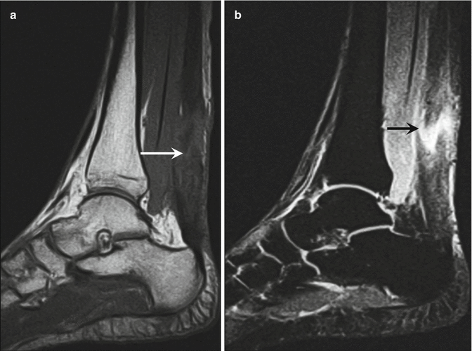
Fig. 22.7
Grade 3 strain of the Achilles tendon. (a) Sagittal T1-weighted MRI of the ankle shows marked thickening of the Achilles tendon (white arrow) and disrupted surface contours both anteriorly and posteriorly. (b) Sagittal STIR MRI shows intense high-signal-intensity fluid in between the frayed free ends of the tendon proper and the musculotendinous junction (black arrow) indicative of complete fiber disruption with edema and hemorrhage
22.2.3 Osseous Injuries
Weight-lifting injuries can result in a number of osseous injuries depending on the mechanism of injury. Accidental injuries cause distal extremity fractures from dropped weights on the foot or pinching of the fingers (Myer et al. 2009). Injuries caused by a cumulative load can produce overuse syndromes and stress fractures, while injuries related to acute overloading can produce avulsion fractures, macro-fractures, and joint dislocations (Reeves et al. 1998a, b; Anderson 2006; Rowe 1979). Adolescent lifters have a higher incidence of osseous injuries than their adult counterparts, particularly in the spine and pelvis (Brady et al. 1982).
A common overuse disorder caused by chronic, cumulative overloading of the shoulder is nontraumatic clavicular osteolysis. This disorder is the result of repeated episodes of minor trauma to the acromioclavicular joint which produces osteolysis of the distal clavicle (Schwarzkopf et al. 2008; de la Puente et al. 1999). This painful condition is characterized by widening of the interval between the clavicle and the acromion process owing to bone lysis of the distal clavicle, which usually begins about 6–8 weeks after the initial insult. The process is self-limiting if the lifter stops performing exercises that target the upper extremity and upper torso until symptoms resolve. Occasionally, however, the disorder becomes recalcitrant to conservative treatment and requires surgical resection of the distal clavicle (Kaplan and Resnick 1986; Scavenius and Iversen 1992). The distal clavicular abnormality is clearly depicted on MRI, consisting of edema in the distal clavicle, widening of the acromioclavicular joint, and articular erosions extending into the subchondral region of the bone (Fig. 22.8) (Yu et al. 2000).
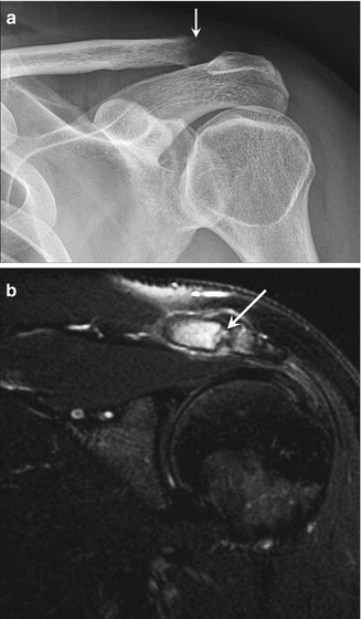
Fig. 22.8
Distal clavicular osteolysis. (a) Frontal radiograph of the left shoulder shows marked osteopenia in the distal clavicle (arrow) and widening of the acromioclavicular joint. (b) Coronal fat-suppressed coronal T2-weighted MRI shows intense bone marrow edema in the distal 2 cm of the clavicle and erosive changes in the articular surface corresponding to an insufficiency fracture (arrow)
Repeated trauma can also lead to periostitis and stress fractures (Haupt 2001). Heavy forearm exercises, for instance, may cause periostitis of the ulna, referred to as forearm splints—analogous to shin splints—leading to stress fractures as the load increases (Wadhwa et al. 1997; Steunebrink et al. 2008). Stress fractures of the ulna have been reported with heavy biceps preacher curls using a straight barbell, an exercise performed with the upper arm resting on an inclined bench while the elbow is flexed against resistance (Hamilton 1984). Intense acceleration in weight loading has been associated with stress fractures of the clavicle and the ribs (Fig. 22.9) (Van der Wall et al. 1999). Fractures of the distal radius and ulna have been described in adolescent weight lifters who have performed exercises requiring a barbell to be lifted above the head (Gumbs et al. 1982). The two contributing factors identified when fractures occur are maximal weight and a loss of balance that causes the barbell to drift back behind the head so that the wrists become hyperextended.
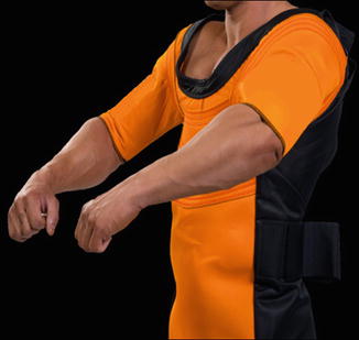
Fig. 22.9
Bench shirt. The apparatus is used during the bench press and is composed of tight nylon so that it is capable of stretching. The sleeves are designed to keep the arms internally rotated and adducted so that as the barbell descends toward the chest, potential energy accumulates in the fibers of the shirt and allows for some spring to the upward portion of the lift. For experienced lifters, the bench shirt can increase performance by more than 20 %
A loss of balance and overloading have also been identified as strong contributing factors in the development of acute-onset low back pain. Sudden loss of balance triggers corrective neuromuscular responses with forceful muscle contractions that can overload and injure spinal tissues. Avulsion fractures frequently involve the extensor mechanism of the knee, the biceps, and triceps tendons and the tendinous attachments of the pelvis. They are usually the result of maximal loading during a particular exercise which prevents the completion of the motion. Ultimately, failure occurs at the enthesis (Bach et al. 1987). A squat is an exercise performed with a barbell rested squarely on the upper back while the trunk is lowered against resistance by simultaneous flexion of the hip and knee. Descent stops when the top of the leg is parallel with the floor. Active contraction of the gluteal and hamstring muscles of the posterior thigh and the quadriceps muscles of the anterior thigh then reverses the movement. This motion exerts tremendous stress on the ischial insertion of the hamstring muscles and the quadriceps tendon insertion on either the superior pole of the patella or tibial tubercle and is exacerbated by eccentric contraction (Bowers et al. 2004; Beltran et al. 2012). Cases of complete rupture are rare but have been reported when there is underlying tendinosis, prior surgery, or previous injury (Yu et al. 1994).
22.3 Specific Weight-Lifting Injuries
22.3.1 The Chest
A common core exercise used by strength athletes is the bench press. In this exercise, a person lying supine brings a weighted barbell from a position of full arm extension down to the chest by passive flexion of the elbows and then reverses the motion by pushing the bar against gravity to a position of full arm extension. The bench press motion involves movement in the sagittal (flexion/extension), coronal (abduction/adduction), and transverse (horizontal adduction) planes (Fees et al. 1998). It is an efficient method of increasing upper body strength and power and developing the triceps brachii muscles and chest musculature. The bench press presents a challenge to the lifter because the shoulder girdle is the principal support for the motion of the barbell to and from the chest.
The bench shirt is an apparatus that increases the loading capacity that a lifter is capable of by creating potential energy during the exercise (Fig. 22.9). With the use of a bench shirt, a lifter is capable of performing the bench press with a higher load than without the use of a shirt, and as such, there are more severe injuries such as fractures of the arm. The incidence of pectoralis major muscle tears also has increased, and when the shirt fails or rips during the exercise, a lifter is particularly vulnerable. A recent review of pectoralis major tears and treatment found that 48 % of pectoralis major tears occurred during weight lifting, although this injury can occur in other sports (ElMaraghy and Devereaux 2012). The usual mechanism of injury to the pectoralis major is eccentric shortening under heavy load when performing bench press (Provencher et al. 2010; de Castro et al. 2014). Additionally, there is a reproducible “sticking period” in the bench press exercise that usually begins about 0.2 s after the initial upward movement that lasts on average about 0.9 s caused by diminishing potentiation of the contractile elements of the pectoralis and deltoid muscles (van den Tillaar and Ettema 2010). This is another potentially hazardous part of the lift where injuries to both the chest and upper arm may occur and is independent of whether a lifter is using a barbell or dumbbells (Tillar and Saeterbakken 2012).
22.3.1.1 Relevant Anatomy
It is important to understand the anatomy of the pectoralis muscles to be able to accurately assess injuries to them (Connell et al. 1999, 2000; Lee et al. 2000; Zvijac et al. 2006; Petilon et al. 2005). The pectoralis major muscle is a fan-shaped muscle with three major origins. The smaller clavicular head arises from the anterior surface of the medial two-thirds of the clavicle, while the larger sternal head arises from the anterior surface of the manubrium and sternal body. Some tendinous fibers of this head cross medially with those from the opposite side. The deeper fibers of the sternal head arise from the cartilage of the first six ribs. There is also a small abdominal head that arises from the aponeurosis of the external oblique muscle. The muscle fibers of all heads converge as they approach the proximal humerus and are inseparably blended just proximal to their insertion onto the lateral lip of the bicipital groove of the humerus.
At the humerus, the fibers that originate from the clavicular head lie anterior to the sternal head fibers and blend inferiorly with the tendinous insertion of the deltoid muscle. The upper insertional fibers of the sternal head lie beneath the clavicular fibers and blend with the deep surface of the pectoralis tendon. The lower fibers of the sternal head curve upward with the abdominal head to insert behind the upper sternal fibers. The abdominal fibers have the most superior attachment on the humerus and contribute to the fascial sheath covering the biceps tendon. A bilaminar tendon is produced with the insertional fibers from the clavicular head having the most inferior attachment onto the humerus and the abdominal fibers having the most superior attachment. The superficial lamina is formed by the upper sternal fibers blended with fibers from the clavicular head, and the deep lamina is formed by the lower sternal and abdominal fibers.
The pectoralis minor muscle arises from the second through fifth ribs and inserts on the coracoid process along with the conjoint tendon of the coracobrachialis and short head of the biceps brachii muscles (Kalra and Neri 2010).
22.3.1.2 Injuries of the Pectoralis Major Muscle
The main function of the pectoralis major muscle is adduction, flexion, and internal rotation of the humerus. An injury can occur during the bench press motion when it is associated with forced abduction of the upper arm at the point of maximal eccentric contraction (Kretzler and Richardson 1989; Wolfe et al. 1992). Clinical assessment of this injury is often misleading since hemorrhage, tenderness, and spasm can alter the physical examination. An avulsion of the tendon from the humerus is generally repaired surgically, as are high-grade tears occurring at the distal myotendinous junction. Prompt surgical repair of a complete avulsion prevents atrophy, fibrosis, and muscle scarring from occurring and should be performed within 6 weeks of injury.
MRI is particularly useful in determining the extent of the tear when surgery is contemplated (Miller et al. 1993; Ohashi et al. 1996). The normal tendon of the pectoralis major muscle is best seen on axial images as inserting on the anteromedial surface of the proximal humeral shaft as a thin, low-signal-intensity linear structure located directly anterior to the coracobrachialis muscle body. The tendon is often demarcated by fat on both sides and is situated directly posterior to the deltopectoral groove. The length of the tendon varies from 5 to 15 mm before insertion in the humerus, and the width of the insertion varies between 4 and 6 cm (Fig. 22.10). The superior edge of the pectoralis major tendon insertion is at the level of or within 1–1.5 cm inferior to the quadrilateral space and approximately 5–10 mm superior to where the lateral head of the triceps muscle is first seen on the axial images.
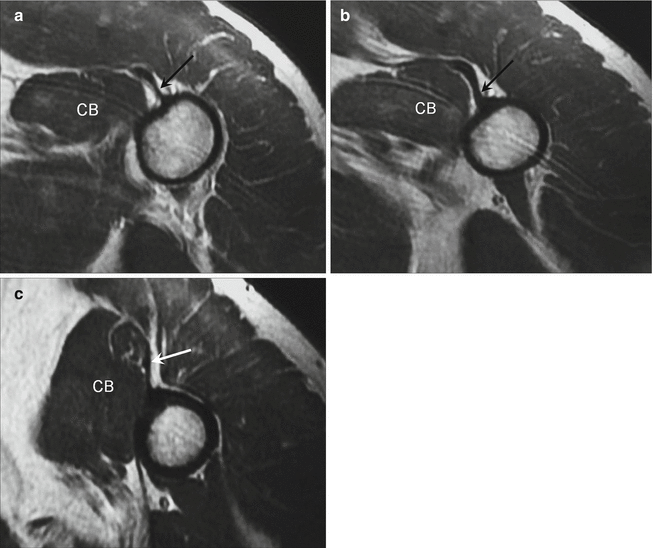
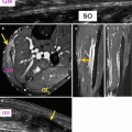
Fig. 22.10
Pectoralis major tendon-bone junction. (a–c) Sequential axial T1-weighted MRI from superior to inferior show the normal appearance of the insertion of the pectoralis major tendon (arrow). Note that the tendon thickens (b) where it is bilaminar. The coracobrachialis muscle (CB) is a reliable landmark for identifying the tendon. The musculotendinous junction is also depicted
The classification of tears depends on the degree (complete or partial) and the location (enthesis, myotendinous junction, or intramuscular), but the location of a tear in large part dictates whether surgery should be performed. Expeditious diagnosis has been shown to result in better outcomes. The majority of pectoralis major muscle injuries are partial tears at the myotendinous junction, but those that tend to be treated are generally more severe injuries (Connell et al. 1999). Complete avulsions from the humerus occur in about 20 % of cases. Complete tears can involve only one head, and these injuries have a better outcome.
Acute tears demonstrate high signal intensity at the rupture on fluid-sensitive sequences usually with retraction of the tendon so that a discernible gap is visible at the enthesis (Fig. 22.11). The tear may be associated with a hematoma and/or periosteal stripping (ElMaraghy and Devereaux 2012; Connell et al. 1999). Most complete tears affect both the clavicular and sternal heads and may be associated with displacement of the biceps tendon from its normal position within the intertubercular groove as well. Early surgical repair of tears at the enthesis has been shown to yield a better outcome for athletes; therefore, immediate diagnosis is advocated. It also is associated with a better cosmetic outcome. High-grade or complete tears at the distal myotendinous junction are also treated surgically when acute, i.e., within 6 weeks of injury (Fig. 22.12). Low-grade partial or intramuscular tears have good outcomes with conservative management (Fig. 22.13). The treatment of chronic tears, those older than 6 weeks from the initial injury, is considered controversial. Some patients who have been treated with a surgical repair have reported a worse outcome. Still, some clinicians advocate delayed repair in high-caliber athletes utilizing a larger incision (which allows better tendon mobilization) since varying degrees of success returning to a competitive level have been reported.
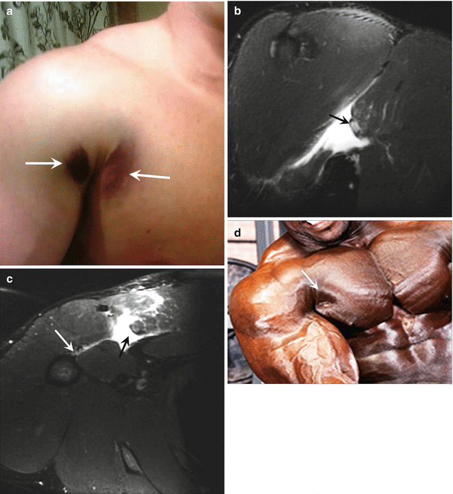
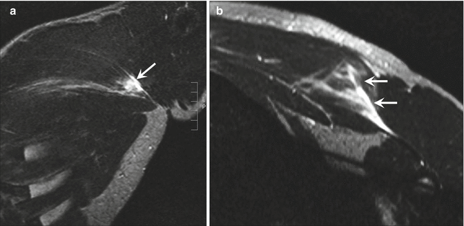
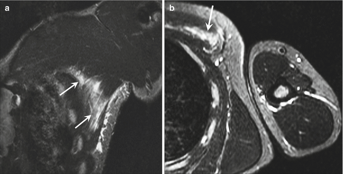
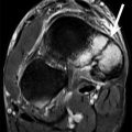
Fig. 22.11
Pectoralis major muscle tear at the enthesis. (a) Characteristic bruising pattern in upper arm and axilla is shown in a patient with a complete avulsion of the pectoralis major muscle (arrows). (b) Coronal fat-suppressed T2-weighted MRI of the chest shows complete avulsion of the clavicular and sternal heads of the right pectoralis major muscle at the tendon-bone junction (arrow). (c) Axial fat-suppressed T2-weighted MRI shows that the muscles are retracted resulting in a gap between the muscle and its insertion (black arrow). Note the absence of the humeral attachment (white arrow). (d) When chronic, it results in a dimple-like defect (arrow)
Fig. 22.12
Pectoralis major muscle tear at the musculotendinous junction. (a) Coronal fat-suppressed T2-weighted MRI shows a focal area of interstitial edema in the lateral aspect of the clavicular head of the pectoralis major muscle (arrow). (b) Axial fat-suppressed T2-weighted MRI shows that the tear (arrows) is located within the musculotendinous junction
Fig. 22.13
Pectoralis major muscle intramuscular tear. (a) Coronal fat-suppressed T2-weighted MRI shows interstitial edema in the inferior sternal and the abdominal heads of the pectoralis major muscle (arrows). Overlying subcutaneous edema corresponded to skin ecchymosis. (b) Axial T2-weighted MRI shows that the tear is intramuscular (arrow)
Partial tears, which constitute the majority of pectoralis muscle injuries, involve the myotendinous junction and less frequently involve the muscle body (Carrino et al. 2000; Lee et al. 2000). The total myotendinous length varies with the each head, measuring 8.5–16 cm for the clavicular head, 13–17 cm for the sternal head, and 14.5–15.5 cm for the abdominal head. Acutely, hemorrhage produces interstitial T2 signal changes in the myotendinous junction with focal fiber disruption and T1 hyperintensity reflecting extracellular methemoglobin. Partial tears also may occur within the muscle itself or along the medial sternal attachment. Grade 1 tears have a feathery appearance and typically involve a small area of the muscle. Larger tears disrupt the architecture of the muscle and may be associated with a hematoma and hemorrhagic fluid deposition in between adjacent muscle interfaces and along the fascia. Patients with low-grade partial tears generally do well with conservative treatment, although return to full strength often requires lengthy rehabilitation and may not be completely possible when the initial tears are associated with large hematomas. High-grade partial tears tend to be surgically repaired in our institution.
22.3.1.3 Injuries of the Pectoralis Minor Muscle
Isolated pectoralis minor tendon tears are rare with only a few cases reported in the literature, all occurring in athletes in contact sports (Kalra and Neri 2010; Li et al. 2012). The main function of this muscle is stabilization of the scapula by drawing it inferiorly and anteriorly against the thoracic wall; therefore, rupture is likely related to sudden muscle contracture to maintain scapular position during an impaction injury.
On MRI, the characteristic appearance is deformity and edema of the pectoralis minor tendon insertion on the coracoid process associated with hyperintense T2 signal intensity related to interstitial edema within the fibers of the pectoralis muscle in a patient with a partial tear. When the tendon is avulsed, a fluid-filled gap is produced from retraction of the muscle, as well as deposition of hemorrhagic fluid along the anterior and posterior epimysial margins (Kalra and Neri 2010).
22.3.1.4 Stress Fractures in the Chest
Accidental trauma may occur during the bench press exercise resulting in trauma to the chest, neck, or face. Mishaps in the exchange of the barbell during a “hand-off,” when the bar is transferred from the person assisting a lifter to the person doing the exercise, may lead to accidentally dropping the weight onto the chest, neck, or head, causing fractures of the sternum or ribs, breaks in the costal cartilage, pericardial contusions, tracheal transection, facial fractures, and intra-abdominal trauma (Fig. 22.14). An unusual disorder associated with weight lifting is the development of a stress fracture of the manubrium or sternum (Fig. 22.15) (Robertsen et al. 1996; McCurdie et al. 1997). It occurs when there is a discrepancy between rapid development of the anterior chest musculature and the strength of the bones.
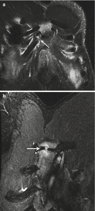
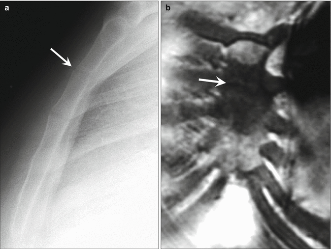
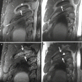
Fig. 22.14
Accidental injuries to the osseous chest. A dropped barbell loaded with weights can produce injuries to the osseous thorax as well as the intrathoracic contents. These different patients presented with a costal cartilage fracture (arrow in a) and a manubriosternal junction contusion (arrow in b) seen in these coronal fat-suppressed T2-weighted MRI
Fig. 22.15
Sternal stress fracture. (a) Lateral radiograph shows a subtle lucency at the synchondrosis between the first and second sternal segments (arrow). (b) Coronal T1-weighted MRI shows intense bone marrow edema on both sides of the stress fracture (arrow)
Another unusual location for a stress fracture in the chest is the rib, which occurs in Olympic-style weight lifters (Eng et al. 2008; Prisk and Hamilton 2008). Patients present with shoulder pain during the act of a “clean and jerk,” where the overhead lift produces stress on the first rib. Bone scintigraphy and CT are optimal imaging modalities for diagnosis when this condition is suspected (Fig. 22.16).
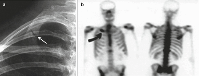
Fig. 22.16
Stress fracture of the first rib in an Olympic weight lifter. (a) Frontal radiograph of the upper right thorax shows a displaced fracture of the right first rib (arrow). (b) Frontal bone scan performed 3 months earlier showed a stress fracture that was misdiagnosed as costochondritis (With permission from Habib et al. (2004))
22.3.2 The Shoulder
Weight-lifting exercises that target the shoulder muscles can place considerable stress across the shoulder, effectively converting this non-weight-bearing joint into one that is weight bearing. The shoulder complex has been alluded to as one of the most prevalent regions of injury accounting for as many as 36 % of weight-lifting injuries (Kolber et al. 2010, 2013). Heavy bench presses, squats, and military presses can strain the trapezius and paracervical musculature as well as the rotator cuff and biceps tendons. In the bench press, the shoulder muscles have to alternate between maximal concentric and eccentric contraction when stabilizing, lifting, or lowering the barbell. A weight lifter who pushes to achieve higher limits of performance and the inherent unfavorable position of the rotator cuff during lifting are two factors that contribute to rotator cuff tears (Neviaser 1991). Repeated trauma to the glenohumeral joint can contribute to labral tears, labrocapsular dysfunction, and resultant shoulder instability, which also may precipitate rotator cuff disease (Yu et al. 2002). In older lifters, the risk for injuries is higher in one-repetition maximum strength testing (“maximal one-lift effort”) than during training, and careful evaluation of all aspects of cardiovascular and musculoskeletal fitness is essential for injury prevention (Sousa et al. 2014).
The biomechanics involved in repeatedly moving from a horizontally adducted to a horizontally abducted position frequently produces compression of the distal clavicle (Fees et al. 1998). The prevalence of nontraumatic osteolysis of the distal clavicle has been reported to be about 28 % in elite weight lifters (Scavenius and Iversen 1992). Bilateral glenohumeral joint dislocations have been reported in lifters using wide-grip benches which cause the glenohumeral joint to act as the fulcrum to the bench press motion (Cresswell and Smith 1998; Heggland and Parker 1997; Maffulli and Mikhail 1990). In most cases of dislocation, fatigue is a major contributing factor. Trends that increase the likelihood of injury include intrinsic risk factors such as joint and muscle imbalances and extrinsic risk factors such as improper technique (Kolber et al. 2010).
Labrocapsular dysfunction may lead to shoulder instability. While the forces that act on the shoulder during a maximal bench press are not sufficient to cause a dislocation, the cumulative force is sufficient to cause subluxation over time. The subluxation can either contribute to the avulsion of the posterior scapular periosteum or may be its end result (Yu et al. 2002; Mair et al. 1998). Conservative measures may be insufficient in relieving symptoms, and surgical debridement and posterior labral repair often are required to reestablish pain-free participation in weight lifting.
22.3.2.1 Relevant Anatomy
The glenohumeral joint of the shoulder is inherently unstable partly because the area of the glenoid fossa is small and in contact with only a small area of the humeral head at any one time. Furthermore, the glenoid joint surface is perpendicularly oriented to the axis of the axial skeleton and the scapula is anteverted providing minimal osseous support inferiorly and anteriorly. Lifting motions that expose an imbalance between the force developed by the periarticular muscles and the actual load of the weight, coupled with fatigue, may predispose the lifter to dislocations and instability.
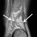
The flat bench press and the inclined/declined variations involve a motion that is particularly risky when overloaded, and there are several reports of bilateral anterior shoulder dislocations in the literature occurring during the exercise. Even when forces acting on the shoulder during a maximal bench press are not sufficient to cause a dislocation, it may be sufficient to cause subluxation particularly over a length of time (Siewe et al. 2011). Chronic posterior subluxation can contribute to the posterior labrocapsular periosteal sleeve avulsion (POLPSA) lesion or a posterior labral tear (Fig. 22.17) (Yu et al. 2002).
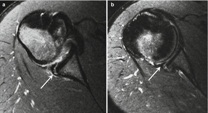
Fig. 22.17
Posterior labral periosteal sleeve avulsion (POLPSA) lesion. (a) Axial fat-suppressed proton density-weighted MRI of the shoulder shows a typical appearance of the POLPSA lesion. The periosteal sleeve formed by the avulsed periosteum tends to remains attached to the posterior glenoid (arrow), creating a redundant recess that communicates with the joint. (b) After 3 months, the posterior displacement of the posterior labrum has increased (arrow)
22.3.2.2 Posterior Labrocapsular Periosteal Sleeve Avulsion
The capsular attachment of the glenohumeral joint and labrum is optimally evaluated with MRI. On MRI, the posterior labrocapsular junction is best depicted on axial images. The posterior labrum is homogeneously hypointense in signal and may appear blunted, rounded, or triangular. The posterior capsule attaches to the base or the apex of the labrum, blending in with the posterior scapular periosteum. A posterior labral tear creates a defect in the labrum or at its base and may be associated with signal changes in the hyaline cartilage of the glenoid fossa. MR arthrography is a useful adjunct to routine MRI since the labrocapsular junction is more accurately assessed when there is joint distention. CT arthrography is recommended if there is a contraindication to MRI.
The POLPSA differs from a posterior labral tear or a posterior Perthes lesion in that, like its anterior counterpart, the posterior periosteum strips off the glenoid rim creating a patulous recess which can then fill in with fibrosis which permanently displaces the labrum (Fig. 22.18) (Yu et al. 2002). Unlike a reverse Bankart lesion, the periosteum/posterior capsule does not rupture. The operative management differs since the periosteal sleeve must be reduced in order to reattach the labrum.
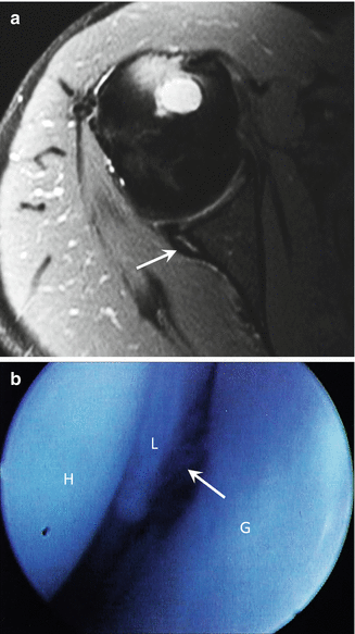
Fig. 22.18
Posterior labrocapsular periosteal sleeve avulsion (POLPSA) lesion. (a) Axial fat-suppressed proton density-weighted MRI of the shoulder shows a posteriorly displaced POLPSA lesion (arrow). (b) Arthroscopic image shows the patulous opening to the periosteal sleeve (arrow). H humerus, L labrum, G glenoid
22.3.3 The Arm
Injuries of the arm are relatively common accounting for about one-fourth of all injuries incurred while weight lifting (Quatman et al. 2009; Amadio 1990). The elbow and wrist are particularly susceptible to acute injuries when resistance training significantly increases stress in the arm, leading to rupture of the biceps or, occasionally, the triceps muscle. Less frequently, a fracture of the radius and ulna or dislocation of the elbow may occur. Elbow dislocations are particularly common in Olympic-style weight lifters during the press phase of their lift when the barbell is pushed above the lifter’s head. Valgus and posterior loading can produce posterior dislocation of the elbow or acute/chronic strains of the ulnar collateral ligament (Bethapudi et al. 2013). Chronic overloading may cause several overuse syndromes including medial and lateral epicondylitis, ulnar neuropathy, tendinosis of the triceps and biceps tendons, and exercise-induced compartment syndrome.
Related posts:

Stay updated, free articles. Join our Telegram channel

Full access? Get Clinical Tree


