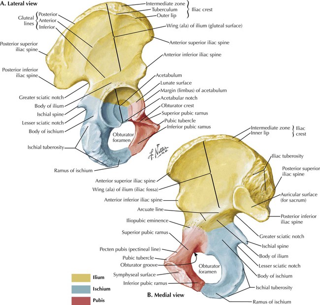
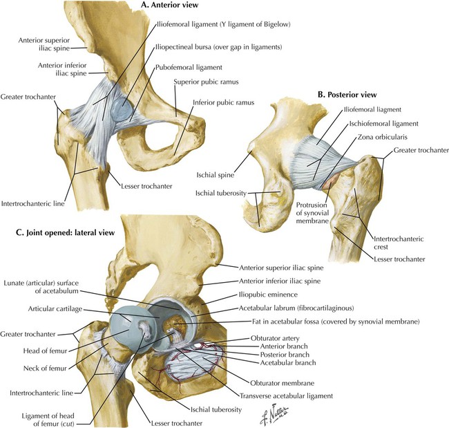
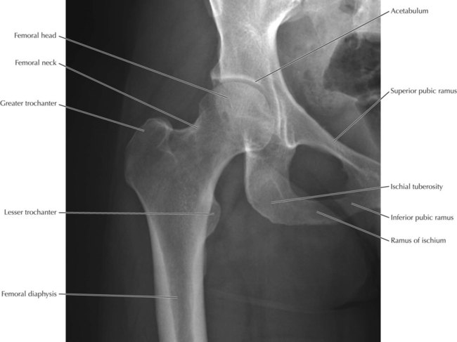
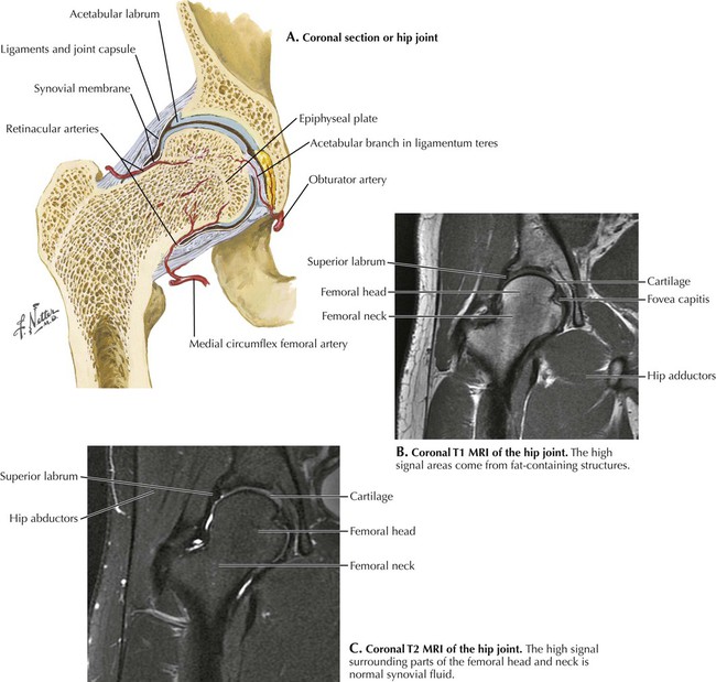
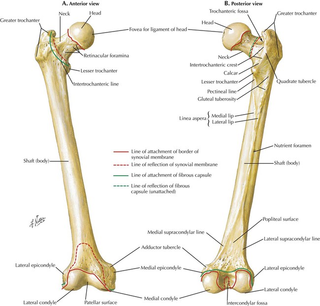
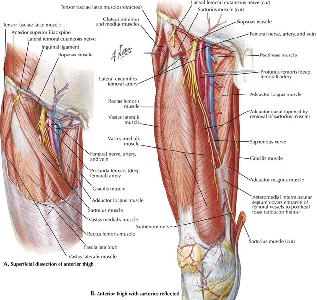
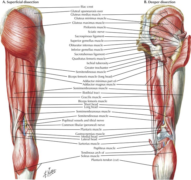
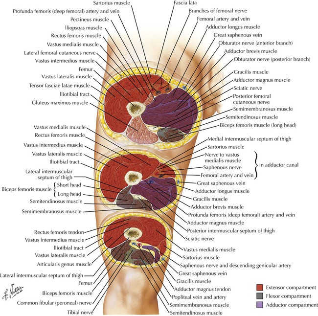
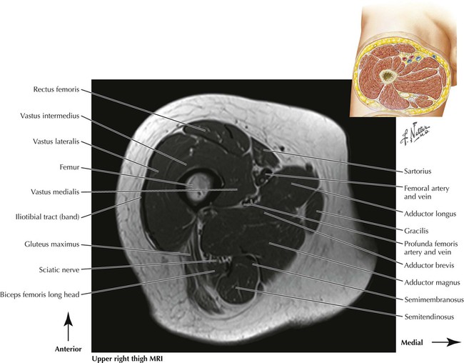
Lower Limbs


Lower Limbs


7.4 Imaging Studies of the Hip Joint


If an initial hip x-ray is normal or inconclusive, magnetic resonance imaging (MRI) is usually the next modality of choice. MRI is advantageous over other imaging modalities in its soft tissue contrast and high resolution. It can often detect pathophysiological changes before they are seen on conventional radiography. It is the most sensitive imaging modality for stress fractures, which appear as a low-signal line on both T1-weighted and T2-weighted images, with a surrounding high-signal area on T2-weighted images representing edema. Fractures of the hip most often occur in the femoral neck or the intertrochanteric region. A potential complication of femoral neck fractures is avascular necrosis (AVN), which can lead to total joint destruction requiring a hip replacement if not caught early. In these two coronal images, there is an area of depression in the otherwise spherical femoral head. This is the fovea capitis, which is the attachment site of the ligamentum teres. It is the only part of the femoral head not covered by articular cartilage.


image


7.9 Upper Right Thigh T1 MRI


On T1 images fat produces a high signal, as seen here in the bone marrow, the subcutaneous tissue, and between muscle fibers and muscle groups. The cortex of the femur is a low-signal area (black). Compare the muscles of the anterior, posterior, and medial compartments with Fig. 7.8. Surrounded by fatty tissue between the anterior and medial compartments are the (superficial) femoral and deep femoral artery and vein. Within the posterior compartment in this image are the semitendinosus muscle, the semimembranosus tendon (low-signal area), the biceps femoris muscle, and the sciatic nerve. Also visualized is the inferior part of the gluteus maximus muscle and the iliotibial tract (area of low signal located lateral to the vastus lateralis). The inferiormost insertion of the gluteus maximus muscle is onto the gluteal tuberosity and the upper extent of the linea aspera.


image

Jan 10, 2016 | Posted by in RADIOGRAPHIC ANATOMY | Comments Off on Lower Limbs

Full access? Get Clinical Tree

Get Clinical Tree app for offline access