Loculated cystic mass with “spiderweb” pattern due to peritoneal adhesions reflecting from ovary
Fine septations throughout collection
Normal ovary at center or lateral margin of cyst
No solid mural or septal nodule to suggest malignancy
Usually anechoic fluid, but can have internal echoes due to hemorrhagic or proteinaceous contents
• MR
Cystic mass with serous fluid (low signal on T1, high signal on T2) and thin internal septations
Margins of cyst outlined by other structures in pelvis (pelvic side walls, uterus, ovaries, loops of bowel)
Morphologically normal ovary at center of cyst
TOP DIFFERENTIAL DIAGNOSES
• Ovarian cancer
• Ovarian cyst or follicle
• Paraovarian cyst
• Hydrosalpinx
• Lymphocele
• Lymphangioma or other congenital cyst
PATHOLOGY
• Most often in women with prior pelvic surgery or inflammatory disorders (endometriosis, pelvic inflammatory disease, inflammatory bowel disease)
CLINICAL ISSUES
• Primarily women of reproductive age
• Can rarely occur in men or post-menopausal women
DIAGNOSTIC CHECKLIST
• Cystic ovarian neoplasm or malignancy if thick septations, solid component/mural nodularity, or large ascites
(Left) Transvaginal US in a patient with history of multiple prior surgeries for Crohn disease shows an anechoic cyst with internal septations enveloping a normal-appearing ovary , a classic appearance for peritoneal inclusion cyst.
(Right) Axial CECT in a patient who had undergone colectomy with creation of a Hartmann pouch shows a loculated, thin-walled pelvic fluid collection partially surrounding the left ovary , characteristic features of a peritoneal inclusion cyst.
(Left) Longitudinal endovaginal US in a woman with 2-year history of pelvic pain and prior surgery for endometriosis demonstrates a complex fluid collection with internal septations surrounding the left ovary , which contains normal follicles. Surgery revealed a peritoneal inclusion cyst and normal ovary.
(Right) Transvaginal US shows the right ovary enveloped by an anechoic fluid collection . A normal follicle is present within the ovary . This is a typical appearance of a peritoneal inclusion cyst.


 Margins of cyst outlined by other structures in pelvis (pelvic side walls, uterus, ovaries, loops of bowel)

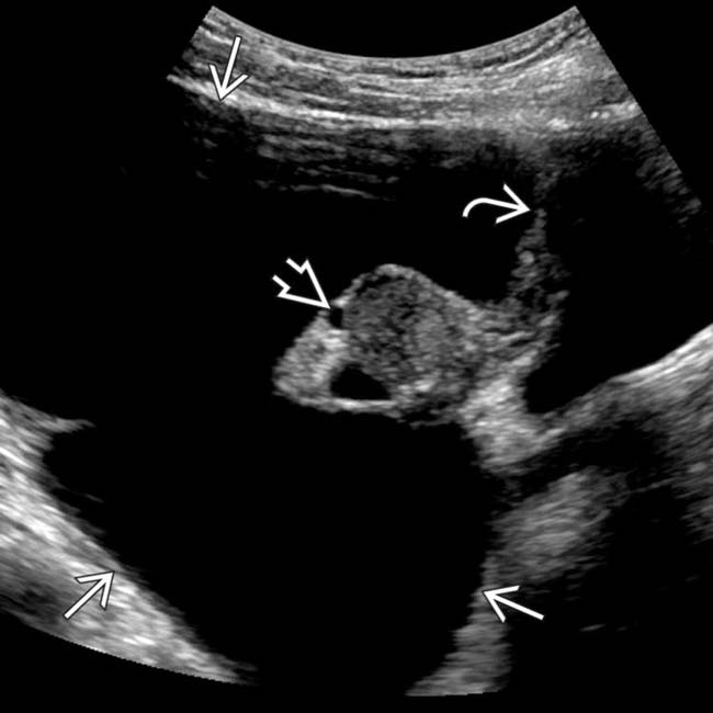
 with internal septations
  enveloping a normal-appearing ovary
  , a classic appearance for peritoneal inclusion cyst.
 shows a loculated, thin-walled pelvic fluid collection
  partially surrounding the left ovary
  , characteristic features of a peritoneal inclusion cyst.
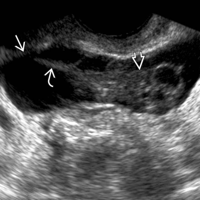
 with internal septations
  surrounding the left ovary
  , which contains normal follicles. Surgery revealed a peritoneal inclusion cyst and normal ovary.
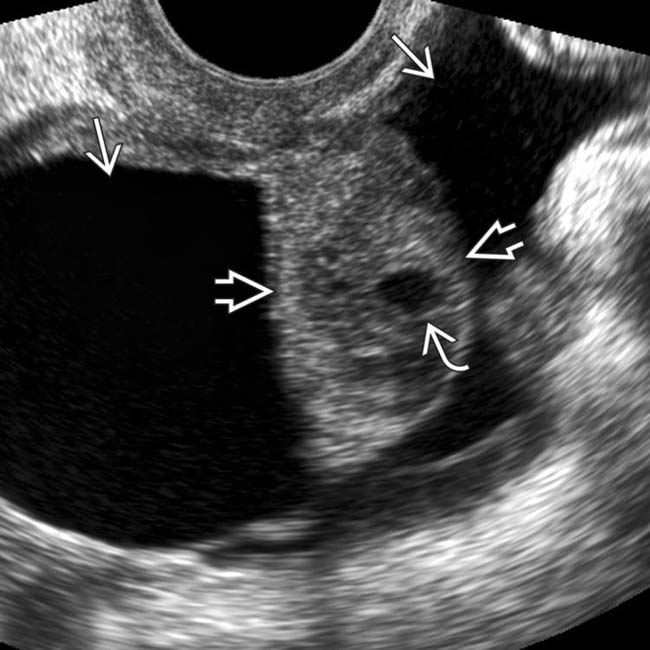
 enveloped by an anechoic fluid collection
  . A normal follicle is present within the ovary
  . This is a typical appearance of a peritoneal inclusion cyst.


 Fine septations throughout collection


