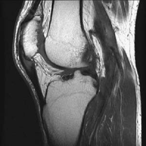
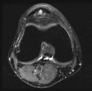
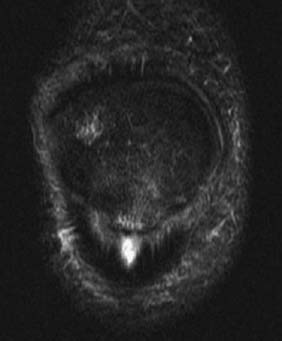
10 Jumper’s Knee

CASE 10


Jumper’s Knee


Anthony G. Ryan and Peter L. Munk


Clinical Presentation


A 20-year-old man presented with anterior knee pain, localized to the lower pole of the patella, focal tenderness at this site on examination, and moderate limitation of active knee extension secondary to pain.


images

Figure 10A


images

Figure 10B


images

Figure 10C


Radiologic Findings


A sagittal T1-weighted image (Fig. 10A) shows focal tendon thickening of the proximal patellar tendon and intratendinous intermediate signal intensity. Focal low signal intensity is shown at the inferior pole of the patella. An axial proton density fat-saturated image (Fig. 10B) shows a focal “cone-shaped” focus of high signal intensity in the proximal patellar tendon. STIR sequence (Fig. 10C) shows a similar “cone-shaped” focus of high signal intensity at the same location. Ill-defined high signal is also demonstrated in the adjacent inferior pole of the patella.


Diagnosis


Jumper’s knee.


Differential Diagnosis


  • Sinding-Larsen-Johansson (SLJ) disease Although frequently used synonymously with jumper’s knee, this term should be reserved for osteochondrosis affecting young adolescents (10 to 14 years). Cerebral spastic children are particularly predisposed. SLJ is caused by persistent traction at the cartilaginous junction of the patella and the patellar ligament, usually at the inferior patellar pole, and usually seen in an active preteen boy who complains of activity-related pain.
  • Infrapatellar bursitis is based on the clinical presentation (easily differentiated from jumper’s knee on ultrasound).
  • Inferior pole sleeve avulsion fracture This entity should be considered in a younger patient.

Discussion


Background


In adults, the weakest link in the muscle-tendon-bone chain is the musculotendinous insertion. In adolescents, prior to epiphyseal fusion, the weakest point is at the tendon-epiphyseal or tendon-apophyseal junction.


Jumper’s knee is the common name given to a partial tear of the patellar insertion of the patellar tendon, most commonly seen in younger athletes, including football players and track and field participants.


Etiology


Patellar tendinosis is thought to be caused by repetitive trauma resulting in microtears at the patellar tendon enthesis, on which background a partial or complete tear may more easily occur. Repetitive violent contractions of the quadriceps occur in “jumping” sports, basketball and volleyball being most commonly implicated.


Extrinsic Factors


  • Hard playing surfaces: cement 38%, parquet 4%, linoleum 23%
  • Length of training sessions
  • Sinding-Larsen-Johansson disease is a condition of adolescence (10 to 14 years). Especially predisposed to this condition are children with cerebral spasticity.
  • A long inferior patellar pole may produce impingement and result in this condition.
  • In the absence of sporting activities, blunt trauma is rarely implicated.

Pathophysiology


There is a spectrum of injury encompassing the following:


  • The changes that occur with shearing of the tendinous fibers from repeated microtrauma can progress to significant degeneration and increase the risk of tendon rupture.
  • Traction with or without contusion and subsequent tendinitis: chronic degeneration eventually disrupts the intratendinous architecture tendon, with a matching increase in pain and activity limitation.
  • In the appropriate age group (i.e., before physeal closure), the patellar component of the lesion is secondary to microavulsions of the epiphyseal cartilage, described by some authorities as evidence that the condition represents a variant of an osteochondrosis affecting the inferior pole of the patella (SLJ).

Clinical Findings

Stay updated, free articles. Join our Telegram channel

Feb 14, 2016 | Posted by in MUSCULOSKELETAL IMAGING | Comments Off on 10 Jumper’s Knee

Full access? Get Clinical Tree

Get Clinical Tree app for offline access