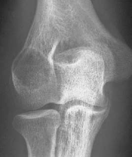
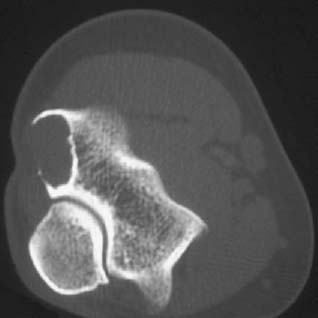
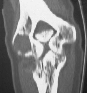
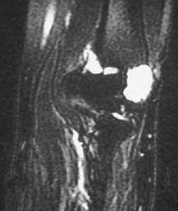
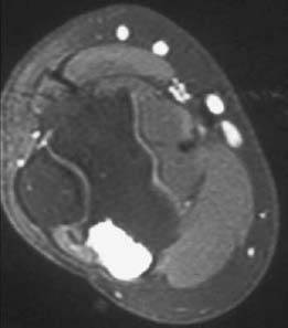
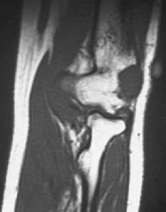
CASE 52 George Nomikos, Anthony G. Ryan, Peter L. Munk, and Mark Murphey A 31-year-old woman presented with increasing left elbow pain that began 3 to 4 years ago, now persisting at rest. Figure 52A Figure 52B Figure 52C Figure 52E Figure 52F An anteroposterior radiograph of the left elbow shows a mildly expansile lytic lesion arising in the lateral epicondyle of the humerus. The lesion demonstrates a narrow zone of transition with a partially sclerotic rim and no mineralized matrix (Fig. 52A). An axial CT scan and a coronal reformatted image (Figs. 52B, 52C) through the lesion show expansion and marked thinning of the overlying cortical bone. The inner border of the lesion is more indolent in appearance and reveals a thin sclerotic rim. The lesion is similar to skeletal muscle in attenuation and does not demonstrate matrix mineralization. The lesion is also similar to skeletal muscle in signal intensity on the T1-weighted MRI (Fig. 52D) and demonstrates very high signal, similar to fluid, on the fat-suppressed T2-weighted image (Fig. 52E). It demonstrates marked diffuse enhancement on the fat-suppressed T1-weighted image (Fig. 52F) after intravenous gadolinium administration. Chondromyxoid fibroma (CMF) of bone.
Chondromyxoid Fibroma of Bone
Clinical Presentation
Radiologic Findings
Diagnosis
Differential Diagnosis
Related posts:
Stay updated, free articles. Join our Telegram channel

Full access? Get Clinical Tree


