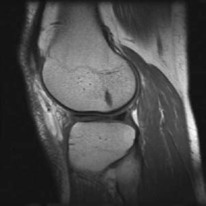
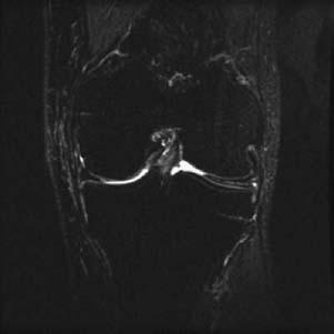
CASE 11 Anthony G. Ryan and Peter L. Munk An 18-year-old girl presented with intermittent locking of her knee after gymnastics training. Figure 11A Figure 11B A sagittal T1-weighted image (Fig. 11A) through the lateral aspect of the knee joint shows intermediate/high signal within the anterior horn of the lateral meniscus, reaching the surface in two locations. The high signal also extends into the posterior horn. The anterior horn is considerably bigger than the posterior horn. A coronal short tau inversion recovery (STIR) image (Fig. 11B) shows intrameniscal high signal intensity across the horizontal width of the visible lateral meniscus. The meniscus extends more centrally than the medial meniscus at the same level. Torn discoid meniscus. None. Discoid meniscus is a relatively common variant of the menisci, present in ~1 in 20 pediatric knees imaged. Discoid menisci are seen far more frequently affecting the lateral meniscus than the medial meniscus, the reported ratio of lateral to medial ranging from 15:1 to 4:1. When present, it is bilateral in 20% of cases. Discoid meniscus is seen more frequently in children and adolescents; thus, some authors suggest it may regress in maturity. The configuration has been presumed to be congenital; however, recent embryologic studies have not upheld this hypothesis, suggesting the lesion is acquired and that discoid menisci may develop when abnormal attachments to the meniscofemoral ligaments of Humphrey and Wrisberg are present (see Wrisberg’s variant below). Discoid meniscus is thought to arise secondary to an abnormal inferior fascicular attachment of the posterior horn, resulting in additional central meniscal growth and subsequent discoid configuration. Morphologically, there is poor constriction of the central portion and thickening of the free end. The abnormal extension centrally predisposes it to trauma between the femoral condyle and the tibial plateau.
Discoid Meniscus
Clinical Presentation
Radiologic Findings
Diagnosis
Differential Diagnosis
Discussion
Background
Etiology
Pathophysiology
Clinical Findings
Related posts:
Stay updated, free articles. Join our Telegram channel

Full access? Get Clinical Tree


