CASE 17 Peter L. Munk, Anthony G. Ryan, and Laurel O. Marchinkow A 23-year-old woman presented with a 1½ -year history of left-sided preauricular pain accentuated by mouth opening and chewing. The patient also reported clicking noises on opening of the mouth. Figures 17A–17C T2-weighted MRIs show the fibrocartilaginous disk to be displaced, such that its posterior band is anterior to the condyle, that is, dislocated (Fig. 17A). On opening the mouth, the condyle has moved anteriorly and regained its normal relationship with the concave undersurface of the disk, which is then said to be “recaptured” (Fig. 17B). The accompanying line diagrams (Fig. 17C) demonstrate the open-mouth view left (the recapture position) and the closed-mouth view right (showing the disk anterior to the head of the condyle). Anterior dislocation of the temporomandibular joint (TMJ) meniscus with reduction on mouth opening. The TMJ is a two-compartment joint, with the segments of the joint separated by a fibrous disk or meniscus. These two compartments do not normally communicate; however, if the disk is damaged or perforated, communication may occur. The meniscus has a donut-shaped elliptical contour and normally travels with the condyle as the mouth opens and the condyle rotates slightly and slides forward over the condylar eminence. Normally, the thin region, known as the intermediate zone, is interposed at the narrowest portion of the joint space between the condylar process and the condylar eminence.
Temporomandibular Joint Disk Dislocation
Clinical Presentation
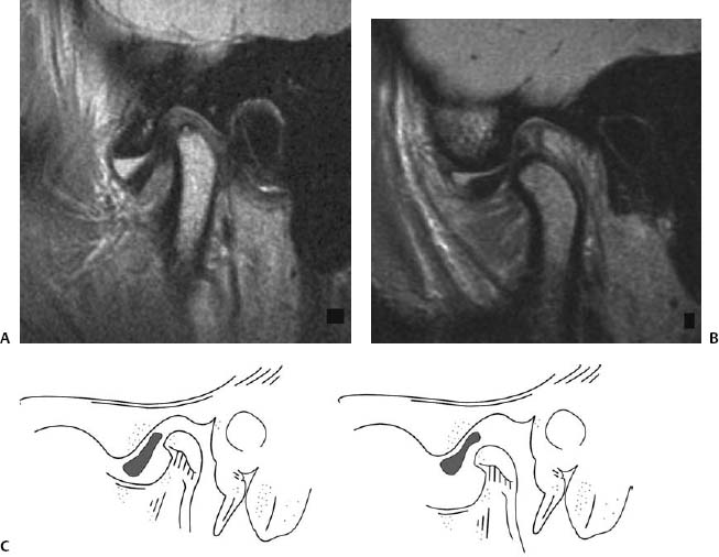
Radiologic Findings
Diagnosis
Discussion
Background
Related posts:
Stay updated, free articles. Join our Telegram channel

Full access? Get Clinical Tree


