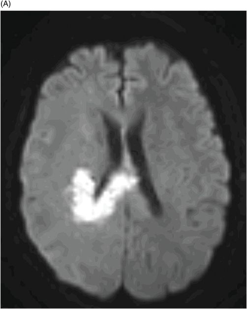
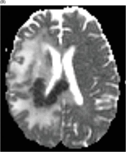
(A) Axial MR DWI and (B) ADC map through the level of the third ventricle (four months post-bevacizumab treatment initiation).
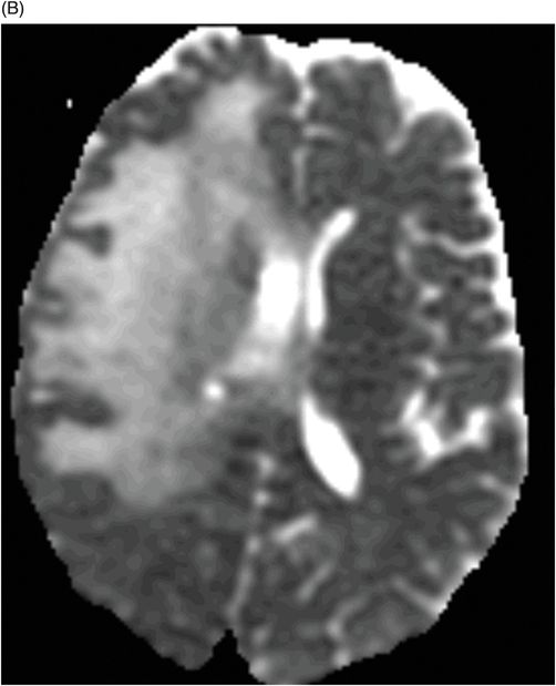
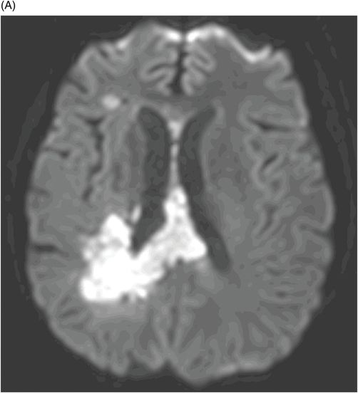
(A) Axial MR DWI and (B) ADC map through the level of the third ventricle (six months post-bevacizumab treatment initiation).
Persistent Diffusion Restriction after Bevacizumab Therapy
Primary Diagnosis
Persistent diffusion restriction after bevacizumab therapy (also known as bevacizumab-related imaging abnormality)
Differential Diagnoses
Recurrence of glioblastoma (GBM)
Acute infarction
Imaging Findings
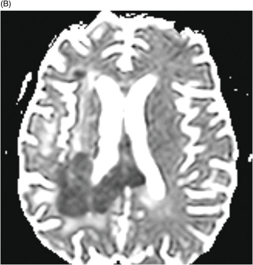
Fig. 105.1: (A) Axial MR DWI and (B) ADC map through the level of the third ventricle two months post-bevacizumab treatment initiation demonstrates a tiny focal area of high signal in the periventricular white matter and in the corpus callosum, without any low ADC value, likely postsurgical changes. Note there is no significant abnormality at the right side of the splenium of the corpus callosum. Also, note the mass effect to the right lateral ventricle. Fig. 105.2: (A) Axial MR DWI and (B) ADC map through the level of the third ventricle four months after initiating bevacizumab treatment demonstrates curved area of diffusion restriction in the right paraventricular white matter reaching the midline through the right side of the splenium of the corpus callosum. Note the very low ADC value in these areas, much lower than expected from recurrence of tumor. Also, note that the mass effect to the right lateral ventricle has significantly improved. Fig. 105.3: (A) Axial MR DWI and (B) ADC map through the level of the third ventricle six months after initiating bevacizumab treatment demonstrates interval worsening of curved area of diffusion restriction in the right periventricular white matter reaching the midline through the right side of the splenium of the corpus callosum. During these six months, however, there was gradual improvement of the peritumoral FLAIR abnormality and no worsening of enhancement (not shown). Relative cerebral blood volume in areas of diffusion restriction showed low value, even lower than normal-appearing white matter. Note a new area of diffusion restriction in the right frontal lobe, an area of distal recurrence.


Stay updated, free articles. Join our Telegram channel

Full access? Get Clinical Tree


