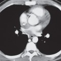
CASE 106 58-year-old woman complaining of progressive shortness of breath Unenhanced chest CT axial (Figs. 106.1A, 106.1B, 106.1C, 106.1D; lung window) and coronal (Figs. 106.1E; lung window) images demonstrate bilateral symmetric ground glass and smooth reticular opacities diffusely throughout the lungs without a zonal predilection. The involved regions of lung parenchyma are sharply demarcated from adjacent normal lung. This combination of ground glass with reticular opacities creates a pattern of disease called “crazy paving.” Note the preservation of lung volume and the absence of lymphadenopathy and pleural effusion. Pulmonary Alveolar Proteinosis • Edema • Infection • Organizing Pneumonia • Neoplasia • Other
 Clinical Presentation
 Radiologic Findings
 Diagnosis
 Differential Diagnosis
 Cardiogenic
 ARDS
ARDS
 Acute Interstitial Pneumonia (AIP)
 Pneumocystis jiroveci Pneumonia
 Viral Pneumonia
 Mycoplasma Pneumonia
 Bacterial Pneumonia
 Bronchiolitis Obliterans Organizing Pneumonia (BOOP)/Cryptogenic Organizing Pneumonia (COP)
 Adenocarcinoma in-situ
 Hemorrhage
 Pulmonary Alveolar Proteinosis
 Sarcoidosis
 Non-Specific Interstitial Pneumonia (NSIP)
 Lipoid Pneumonia
 Subacute Radiation Therapy–Related Pneumonitis (XRT)
 Discussion
Background
Stay updated, free articles. Join our Telegram channel

Full access? Get Clinical Tree


