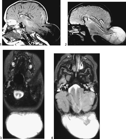
12 Chiari III Malformation

CASE 12


Clinical Presentation


Infant girl presents with dysfunction of the lower cranial nerves, a large, posterior soft mass of the neck present since birth, and microcephaly.


image

Figure 12A


Radiologic Findings


Sagittal MRI T1-weighted image (Fig. 12A1) shows a large posterior herniated sac filled with cerebrospinal fluid and cerebellum as well as posterior parts of the cerebral hemispheres. No subcutaneous fat is seen over it. The bony defect involves the infratentorial and supratentorial occipital bone, and therefore the posterior falx and tentorium as well. This implies that the dural venous sinuses are abnormal. Sagittal MRI T1-weighted image (Fig. 12A2) confirms the same anomalies, together with deformation of the course of the cerebral arteries. The axial T2-weighted images reveal the posterior split in the medulla (Fig. 12A3) and disorganization of the cerebellum (Fig. 12A4). The “heart-shaped” midbrain is tethered posteriorly to the sac (Fig. 12A4).


Diagnosis


Chiari III malformation


Differential Diagnosis


  • Simple occipital meningocele: exceedingly rare, and more usually found at the cervical level. Does not contain any brain tissue, which is clearly shown by MRI. The subcutaneous fat is normal. Treated by simple excision.
  • Occipital dermoid tracts and cysts are more common. The posterior bulging mass often is inflammatory and less elastic. MRI demonstrates the lesion, which is usually extended to the cerebellum, irregular, enhanced, and sometimes infected.
  • Other possible posterior masses of the upper neck include neurofibromas (NF1), hemangiomas, lymphangiomas, and lipomas.

Discussion


Background


  • Chiari III malformation is extremely rare, occurring in as few as 1 per 150 of all Chiari cases, most commonly in girls.
  • Carries the same genetic prognosis as other neural open dysraphisms. In experimental animals, offspring may show any of the following neural closure defects: myelomeningoceles/Chiari II, Chiari III, cranioschizis, exencephaly/anencephaly, rachischizis, and craniorachischizis.
  • On the contrary, Chiari I seems not to belong to the same group of malformations and has no genetic association.

Etiology


Familial/ethnic occurrence similar to Chiari II, commonly associated with maternal dietary folate deficiency or methylene tetrahydrofolate reductase (MTHFR) mutations. Toxins such as Tripterygium wilfordii and arsenic also have been implicated.


Clinical Findings


  • Fetal ultrasonographic or postnatal discovery of occipital mass, often associated with microcephaly. Late diagnosis has been reported to 14 years of age.
  • Soft tissue mass of the upper neck with microcephaly, dystrophic skin, and abnormal hair
  • Brain stem/cerebellar symptoms (cranial nerve deficits, respiratory insufficiency, spasticity, aspiration, and dysphagia)
  • In the older child, symptoms of syncope and headache and signs of raised intracranial pressure may be seen as well as developmental delay.

Complications

Stay updated, free articles. Join our Telegram channel

Dec 21, 2015 | Posted by in PEDIATRIC IMAGING | Comments Off on 12 Chiari III Malformation

Full access? Get Clinical Tree

Get Clinical Tree app for offline access