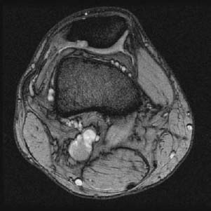
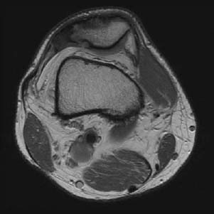
CASE 12 Hema N. Choudur, Anthony G. Ryan, Peter L. Munk, and Laurel O. Marchinkow An elderly woman presented complaining of long-standing anterior knee pain, especially when descending stairs and rising from a chair. Figure 12A Figure 12B Axial MPGR (Fig. 12A) and axial proton density (Fig. 12B) images of the knee reveal a focal increase in signal intensity and a defect in the lateral articular cartilage of the patella with subjacent bony changes, including erosion and sclerosis. Irregularity of the articular surface of the patella with bony erosion of the lateral patellar surface was demonstrated on an accompanying skyline view. Chondromalacia patella. Softening or wearing away of the patellar articular cartilage (chondromalacia patella) causes varying degrees of inflammation and pain. The underlying bone becomes involved when the entire thickness of articular cartilage is destroyed. Severity is graded on a scale from 1 to 4. The patella serves primarily to increase the leverage and therefore the efficiency of the quadriceps muscle. Considerable retropatellar forces are present throughout the entire range of motion of the knee. The articular cartilage is thus compressed against the femoral condyles, exacerbated by acute trauma or chronic overuse. Other anatomical factors causing excessive compression forces are The articular cartilage of the patella consists of collagen fibers oriented tangentially from the articular surface to the subchondral bone; these fibers are disrupted to varying degrees secondary to applied compressive forces. Most of the resulting changes occur at the median ridge of the patella, where the cartilage is thickest. Associated microfractures and sclerosis of the subchondral bone occur in association. Pain along the anterior aspect of the knee while walking, running, or jumping is the usual complaint. It is often aggravated while descending or ascending stairs. Swelling of the knee with crepitus occurs with chronicity.
Chondromalacia Patellae
Clinical Presentation


Radiologic Findings
Diagnosis
Differential Diagnosis
Discussion
Background
Etiology
Pathophysiology
Clinical Findings
Stay updated, free articles. Join our Telegram channel

Full access? Get Clinical Tree


