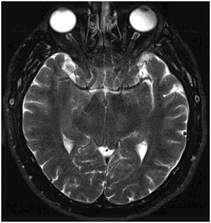
Sagittal T1WI MR at the level of the midbrain.

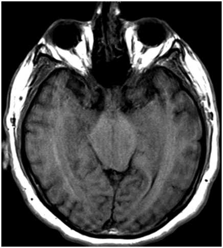
Axial T2WI MR at the level of the midbrain.

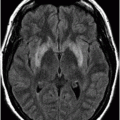
Axial T1WI MR at the level of the midbrain.
Intracranial Hypotension with Midbrain Swelling
Primary Diagnosis
Intracranial hypotension with midbrain swelling
Differential Diagnoses
Midbrain tumor
Abnormality of the diencephalic-mesencephalic junction
Imaging Findings
Fig. 124.1: Sagittal T1WI through the midline demonstrated apparent swelling of the mesodiencephalic junction with complete obscuration of the pontomesencephalic junction, flattening of the anterior surface of the pons, and decreased mammillopontine distance. Transforaminal herniation of the cerebellar tonsils was also noted. Fig. 124.2: Axial T2WI through the diencephalon-mesocephalon junction demonstrated apparent swelling of the mesodiencephalic junction, without any obvious signal abnormality. Note that the third ventricle was compressed severely, with no identifiable CSF signal. Fig. 124.3: Axial T1WI, from a slightly lower level, demonstrated swollen appearance of the mesodiencephalic junction. Note that obscuration of the interpeduncular cistern prevents identification of the cerebral peduncles.
Discussion
Postural headache is a typical manifestation of spontaneous intracranial hypotension (SIH). The imaging features suggestive of intracranial hypotension with midbrain swelling (IHMS) in this patient include severe brain sagging with transtentorial descent of the third ventricle and the diencephalon, swollen appearance of the diencephalon, flattening of the anterior pons, and a narrow pontomesencephalic angle.
Although a tumor-like appearance of the mesencephalic-diencephalic junction is noted, no abnormal T2 signal, enhancement, or diffusion restriction suggestive of tumor was seen. Similar patient imaging features have been described in the literature as abnormalities of the diencephalic-mesencephalic junction or congenital malformations. However, doubts about the existence of such entities persist.
Spontaneous intracranial hypotension is a headache disorder defined by presence of a postural headache associated with any of the following: photophobia, nausea, hyperacusis, tinnitus, neck stiffness, and the presence of low CSF pressure on MRI with or without imaging evidence of an active CSF leak that responds to blood patching. Spontaneous intracranial hypotension typically presents around 40–60 years of age as a severe orthostatic headache (worsens in an upright position and improves in a supine position). It is a relatively rare disorder with an estimated annual incidence of 5 cases per 100,000. There is a slight female-to-male predominance with a 2:1 ratio.
The etiology of SIH is ascribed to a decrease in CSF pressure, which current evidence suggests is most commonly a result of a spontaneous spinal CSF leak. The cause of the spontaneous leaks is unclear, but heritable connective tissue disorders that may cause dural weakness, such as Marfan syndrome and Ehlers-Danlos syndrome type II, have been implicated. Significant CSF leaks are usually located in the spine, most often at the thoracic level. Reduced CSF pressure may also be a result of trauma, CSF overshunting, exercise, violent coughing, lumbar puncture, severe dehydration, or spontaneous dural tear from a ruptured arachnoid diverticulum. In one-third of cases, a history of minor trauma precedes symptomatic onset. Rarely, spinal pathology, such as degenerative osteophytes, can lead to piercing of the dura. The pathophysiology is explained by the Monroe-Kellie hypothesis, which indicates that intracranial volume is constant, being a sum of brain parenchyma, CSF volume, and intravascular volume. Thus, CSF hypovolemia is compensated by increased intravascular volume and compliant vessel dilatation. Headache could result from stretching of pain-sensitive intracranial structures in a sagging brain, or from vascular congestion.
Typical imaging features of SIH include pachymeningeal thickening and enhancement in both supra- and infratentorial compartments, due to dilated subdural blood vessels. Subdural hygromas due to compensatory fluid accumulation from spinal loss of CSF and subdural hematomas due to ruptured bridging veins are common. These may be seen in 17–60% of patients and are a late finding, usually not occurring without dural enhancement. Magnetic resonance imaging of the brain may be normal in up to 20% of patients. The optic chiasm and hypothalamus drape over the sella as the suprasellar cistern gets crowded or effaced. Pituitary enlargement and congestion may be present. Brain descent is seen in 40–50% of cases, with caudal displacement of tonsils in 25–75%. Angiography may demonstrate diffuse enlargement of cortical and medullary veins. Radionuclide cisternography may detect focal accumulation of radiotracer at the leakage site and poor migration of tracer over convexities, with early detection in the bladder and kidneys. Lumbar puncture, performed if neuroimaging is inconclusive, typically has an opening CSF pressure below 6 cm H2O. Cerebrospinal fluid analysis may show reactive mildly elevated protein levels, and pleocytosis (up to 50 cells/mm3). Computed tomography or MR myelography with contrast may identify the site of the leak, which may be especially useful when planning intervention.
Intracranial hypotension with midbrain swelling is a special variant of SIH in which there is severe brain sagging with transtentorial descent of the third ventricle and the diencephalon, and swollen appearance of the diencephalon: flattening of the anterior pons narrows the pontomesencephalic angle. Unlike other cases of SIH, there is also narrowing of the angle between the vein of Galen and straight sinus. In IHMS, dural congestion, enhancement, and subdural fluid accumulation are absent. The third ventricle is usually extremely thin (slit-like) and is located at the level of the midbrain, compared to its normal position. This swelling is likely due to mild vasogenic edema secondary to functional stenosis of the vein of Galen–straight sinus confluence.
Management of SIH is empirical and no treatment options have been evaluated by randomized controlled trials. Spontaneous remission is common. Conservative management includes initial strict bed rest, analgesics, and adequate oral hydration with caffeine intake. Theophylline and steroids may be considered. If conservative management fails, an epidural blood patch is recommended which involves injecting a volume of 10–20 ml of autologous blood into the epidural space, thought to plug the dural tear, forming a scar in two to three weeks.
Stay updated, free articles. Join our Telegram channel

Full access? Get Clinical Tree


