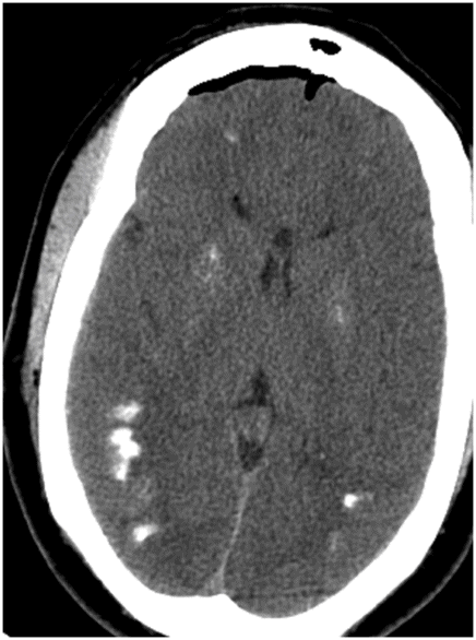
Axial non-contrast CT scan of head through the level of centrum semiovale.
Mineralizing Microangiopathy
Primary Diagnosis
Mineralizing microangiopathy
Differential Diagnoses
Familial idiopathic basal ganglia calcification (FIBGC)
Abnormal calcium metabolism
TORCH infection
Imaging Findings
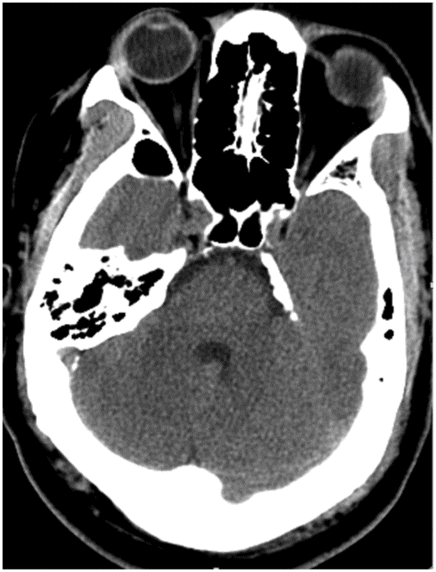
Fig. 129.1: Axial non-contrast CT scan of head through the level of centrum semiovale demonstrated bilateral, chunky calcification at the gray-white junction. Fig. 129.2: Axial non-contrast CT scan of head through the level of basal ganglia demonstrated similar chunky calcification at the gray-white junction, predominantly in the posterior aspect of the right temporal lobe. Faint calcification is also noted in bilateral basal ganglia. Note tiny right frontal pneumocephalus from the maxillofacial trauma. Fig. 129.3: Axial non-contrast CT scan of head through the posterior fossa demonstrated no identifiable calcification in the dentate nuclei.
Stay updated, free articles. Join our Telegram channel

Full access? Get Clinical Tree


