Hippocampal Sclerosis Dementia
Primary Diagnosis
Hippocampal sclerosis dementia
Differential Diagnoses
Alzheimer disease
Vascular dementia
Frontotemporal lobar degeneration
Imaging Findings
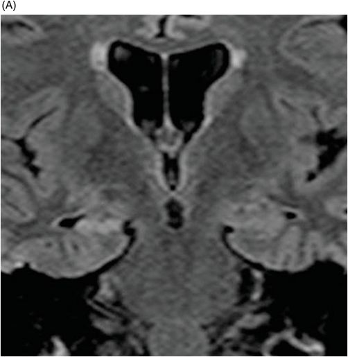
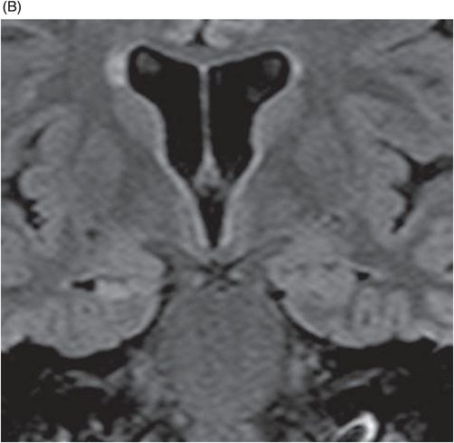
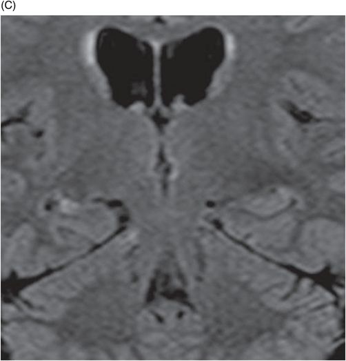
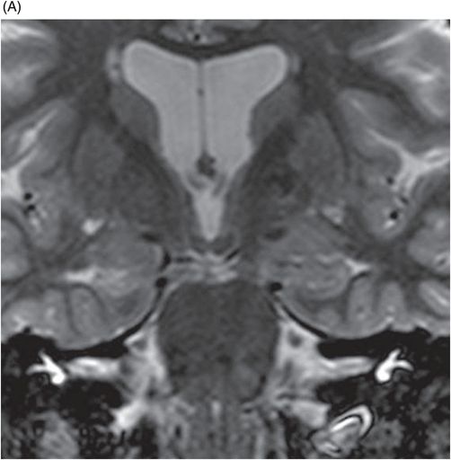
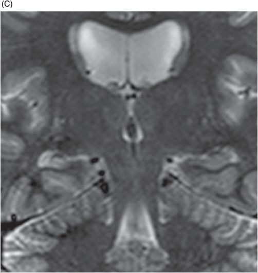
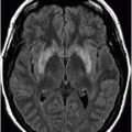
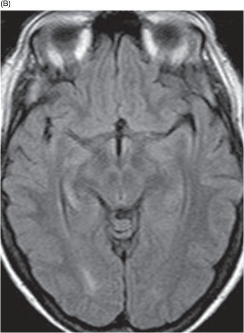
Fig. 13.1: (A–B) Axial FLAIR MR images showed hyperintense signal in the right hippocampus. Fig. 13.2: (A–C) Coronal FLAIR MR images showed isolated hypersignal, as well as signs of marked atrophy on the right hippocampus (head, body, and tail). Fig. 13.3: (A–C) Coronal T2 STIR also showed hypersignal of the right hippocampus, more clearly demonstrating hippocampal atrophy.
Discussion
Pure hippocampal sclerosis (HS) is a rare cause of dementia whose name is derived from a constellation of associated neuropathologic findings. Hippocampal sclerotic neuropathology is characterized by severe neuronal loss and gliosis in the cornu ammonis area 1 (CA1) region of the hippocampus and subiculum. However, the associated findings of other dementing illnesses such as Alzheimer disease (AD), vascular dementia (VD), or frontotemporal lobar denegation (FTD), or clinical evidence of syncope, hypotension, or hypoxia, in association with dementia onset are noticeably absent. Thus, correlating clinical and imaging findings in cases of suspected HS is imperative in order to narrow the differential diagnosis. In this patient, the presence of marked hippocampal atrophy in conjunction with confirmed memory loss strongly suggests a diagnosis of HS.
In addition to demential disorders, other neuropathologies are associated with HS. Mesial temporal sclerosis, or HS, is most commonly associated with prolonged seizures in early infancy and cerebral infarcts are associated with HS. However, in these conditions, the clinical symptomatology and age of affected individuals differs, in comparison to the dementia syndromes.
The estimated prevalence of HS among patients with dementia is variable, ranging from 0.4% to 8%, being more frequent in patients 80 years of age or older. The etiology of HS dementia is unknown. Generally, patients with HS present with cognitive disturbances, mimicking AD and/or FTD, making its clinical diagnosis extremely difficult. It has been reported that these patients have a tendency to become progressively demented and that the duration of their illness is shorter, in comparison to patients with AD without HS.
At imaging, unilateral or bilateral hippocampal atrophy in conjunction with increased hippocampal signal intensity on FLAIR and T2-weighted MR images constitutes the prominent findings.
Stay updated, free articles. Join our Telegram channel

Full access? Get Clinical Tree


