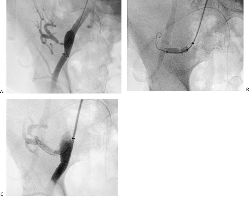
CASE 13 A 47-year-old male who had received a kidney transplant 1 year earlier presented with refractory hypertension. Figure 13-1 Transplant renal artery angioplasty. (A) Digital subtraction angiogram shows high-grade stenosis of transplant renal artery. (B) Fluoroscopic image shows balloon dilation of stenosis. (C) Final digital subtraction angiogram shows alleviation of the stenosis. Renal ultrasound showed a velocity of 350 cm/s in the transplant renal artery. The left common femoral artery was catheterized using the Seldinger technique and a 5-French (F) sheath was inserted. The contralateral common iliac artery was catheterized over the aortic bifurcation with a 5F Rosch Inferior Mesenteric catheter (Cook, Bloomington, Indiana). A right pelvic angiogram was performed revealing a high-grade stenosis of the transplant renal artery (Fig. 13-1A). High-grade stenosis of the transplant renal artery. Five thousand units of heparin were administered intra-arterially. The renal artery was catheterized over the bifurcation using a 5F (Cook, Bloomington, Indiana) Inferior Mesenteric catheter and a Bentson guidewire. The catheter was then exchanged over a Rosen guidewire for a 6 mm- × 2 cm-long low-profile angioplasty balloon catheter. The stenosis was dilated at 6 to 8 atmospheres for 30 seconds (Fig. 13-1B). Follow-up arteriography (Fig. 13-1C) showed no residual stenosis with rapid antegrade flow to the kidney transplant.
Clinical Presentation
Radiologic Studies
Doppler Ultrasound
Renal Angiography
Diagnosis
Treatment
Equipment
Discussion
Background
Related posts:


Stay updated, free articles. Join our Telegram channel

Full access? Get Clinical Tree


