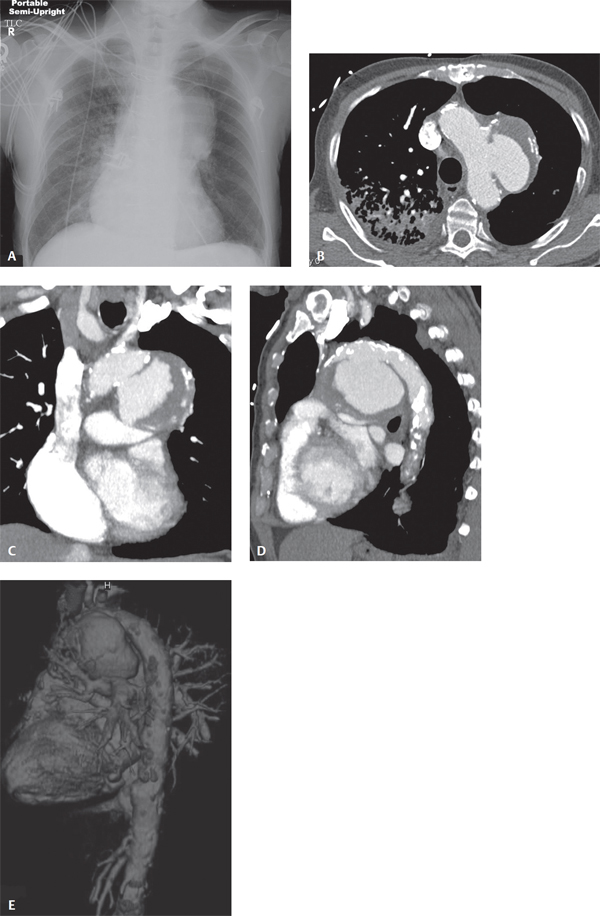
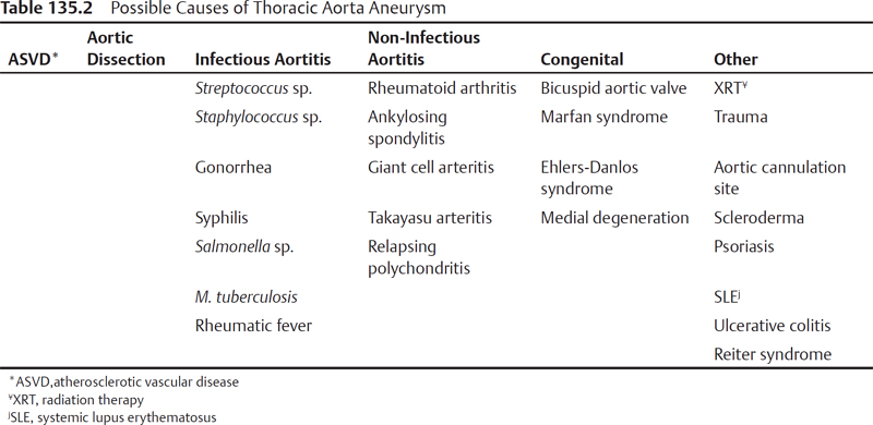
CASE 135 67-year-old man with a history of remote CABG presents with increasing substernal and chest pain AP chest radiograph (Fig. 135.1A) shows an 8.0 cm left-sided focal convex mediastinal mass overlying the aortic pulmonary window contiguous with and in close proximity to the thoracic aorta. Remote CABG surgical changes are evident. Patchy air space disease is seen in the right upper lobe. MDCTA axial (Fig. 135.1B), coronal (Fig. 135.1C), sagittal (Fig. 135.1D), and 3-D color volume-rendered (Fig. 135.1E) images confirm the mediastinal mass is a large saccular aneurysm off the transverse aorta. Note the abundant mural thrombus, displaced intimal calcifications, and extensive atheromatous plaque in the aorta. Right upper lobe air space disease is also seen. Thoracic Aorta Aneurysm • Saccular Aneurysm Sequelae of Ruptured Penetrating Ulcer or Plaque • Saccular Aneurysm Sequelae of Remote Blunt Trauma/Missed ATAI (Fig. 100.13) • Coronary Artery Bypass Graft Aneurysm • Other Non-Vascular Mediastinal Masses (based on chest radiography alone) Dimensions of the normal thoracic aorta at various anatomic regions are summarized in Table 135.1. An aneurysm is a focal, abnormal, irreversible dilatation of a vessel greater than 50% of that of the adjacent normal vessel lumen. Thoracic aortic aneurysms (TAAs) may be classified as either true aneurysms (contain all three layers of aortic wall) or false aneurysms (pseudoaneurysms) (contained only by adventitial or periadventitial tissues). True aneurysms are usually associated with fusiform dilatation of the aorta (80%) and are most commonly due to atherosclerosis. False aneurysms are typically saccular and are most commonly due to trauma, penetrating atherosclerotic ulcers, or infection (Figs. 135.1A, 135.1B, 135.1C, 135.1D, 135.1E). Seventy percent of TAAs result from atherosclerosis and most involve the descending aorta. Other possible causes of TAAs are listed in Table 135.2. Non-syphilitic infection of the arterial wall with subsequent aneurysm formation is called a mycotic aneurysm. Mycotic aneurysms are usually saccular, contain eccentric thrombus, and have a propensity to involve the ascending aorta. Table 135.1 Normal Thoracic Aorta Dimensions Anatomic Location Definition Average Diameter Aortic root Portion of ascending aorta containing aortic valve, annulus, sinuses of Valsalva 37 ± 3 mm Ascending aorta Extends from aortic root to right brachiocephalic artery origin 31 ± 4 mm Arch Extends from right brachiocephalic artery to ligamentum arteriosum attachment 26 ± 4 mm Descending aorta Extends from ligamentum arteriosum to aortic hiatus at the diaphragm 24 ± 3 mm Most patients with TAAs are asymptomatic. Symptoms usually result from compression of adjacent mediastinal structures and may include SVC syndrome; stridor or dyspnea; dysphagia; and hoarseness from recurrent laryngeal nerve compression. Twenty-five percent of patients may complain of substernal chest, shoulder, or back pain. • Contour abnormality and tortuosity of aorta (Figs. 135.1A, 135.2A
 Clinical Presentation
 Radiologic Findings
 Diagnosis
 Differential Diagnosis
 Discussion
Background
Etiology
Clinical Findings
Imaging
Chest Radiography
Related posts:
![]()
Stay updated, free articles. Join our Telegram channel

Full access? Get Clinical Tree


135 Thoracic Aorta Aneurysm