14 The Growing Skeleton of the Hand
With reference to statistical tables, current skeletal age and expected body height can be determined with sufficient accuracy from radiographs. Both values can be determined from the third month of life by examining a radiograph of the left hand.
Normal Development of the Skeleton of the Hand
Ossification of the skeleton begins in the second fetal month and continues rapidly thereafter. Several centers of ossification develop and rapidly fuse at the diaphyses and metaphyses of the tubular bones. The first of these ossification nuclei appears in the clavicula and the upper and lower jaws. Fetal ossification is completed in the eighth to ninth fetal month of life. Under normal developmental conditions, the shafts of all tubular bones, as well as the epiphyseal nuclei of the distal femur, the proximal tibia, and the round bones (talus and calcaneus) are visible at birth. Ossification of all other epiphyses and round bones does not take place until the postfetal period.
Longitudinal growth of the tubular bones parallels ossification of the epiphyses and apophyses after the fetal period. Both processes are completed with closure of the epiphyses in puberty. Sesamoids appear at about the age of 13 years, but differ individually in the time of their appearance and in their number. In the hand, they first appear in the metacarpophalangeal joints. Somewhat later, the relatively constant sesamoids appear in the interphalangeal joints of the thumb, where they can be found in 94% of individuals on the radial side and in 100% on the ulnar side of the hand. Puberty begins at the time of their appearance.
Besides the increase in the number of epiphyses and growth in height, there is also a change in the shape of the epiphyses, which represents skeletal development, as well as that of the entire organism.
Disturbances in Skeletal Maturation
If general development is disturbed ( Table 14.1 ), changes appear that can also be recognized in skeletal maturation. The measurement methods detailed in the following sections enable determination of skeletal age and recognition of a possible retardation or acceleration.
|
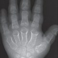
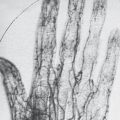
The hand, with its 28 bones (and additional 19 epiphyses), reflects general skeletal maturation ( Fig. 14.1 ). For this reason, a radiograph of the left hand is generally taken to determine skeletal maturation. The radiograph of the hand is considered a reliable indicator for maturation of other body systems and is therefore of special importance in determining the age of a child. The pattern of radioisotope uptake in scintigrams of the hand is also largely dependent on the stage of skeletal development and provides the basis for age-dependent illustrations in tables. Not only bone nuclei but also nonossified cartilage can be demonstrated in MRI. However, these two methods have not yet become part of clinical routine.

Legal and Forensic Considerations
Occasionally determination of the skeletal age is required by the criminal investigation department or courts of law. In most cases, this information is needed to determine the age of asylum seekers or juvenile delinquents, for whom it must quickly be clarified whether juvenile criminal law is still applicable. In most countries, the judicially required degree of certainty is “probability bordering on certainty.” This indicates a probability of over 99%, which is not achievable with a radiograph of the hand alone. Therefore, exact determination of age requires, in addition to a radiograph of the left hand, a physical and dental examination, including a panoramic film of the teeth. The radiologic medical certificate alone constitutes a decision aid for judges and public prosecutors.
Evaluation Methods in Diagnostic Imaging
Age-dependent Factors
Age Determination Up to Three Months
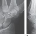
The hands of newborns and infants have neither epiphyseal nor carpal ossification nuclei. Therefore, for determination of the skeletal age in the first three months of life, a radiograph of the lower leg with knee and ankle is taken because the ossification nuclei in the proximal epiphysis of the tibia, the calcaneus, and the talus are physiologically evident at birth. These ossification nuclei can also be demonstrated with ultrasonography in newborns ( Fig. 14.2 ).
Related posts:

Stay updated, free articles. Join our Telegram channel

Full access? Get Clinical Tree


