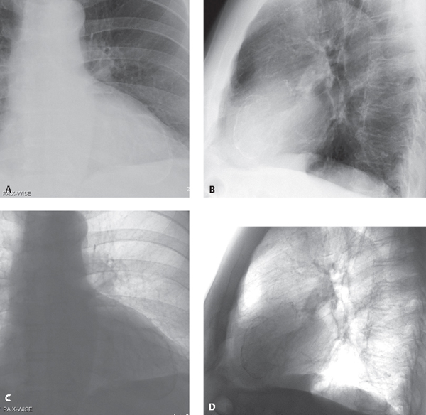
CASE 140 65-year-old man with nonspecific chest discomfort and a remote myocardial infarction in the left anterior descending vascular territory 6 years prior Coned-down PA (Fig. 140.1A) and lateral (Fig. 140.1B) chest radiographs demonstrate ovoid, curvilinear, laminated rings of calcification located more than 2 mm within the outer confines of the cardiac silhouette delineating the anterior and apical walls of the left ventricle. These calcifications are seen to better advantage on the accompanying inverted PA (Fig. 140.1C) and lateral (Fig. 140.1D) images. Fig. 140.1 (Images courtesy of James Messmer, MD, VCU Medical Center, Richmond, Virginia.) Calcified Left Ventricular Aneurysm • Myocardial Calcifications • Pericardial Calcifications (calcific pericarditis) (see Case 141) – Coxsackievirus – Influenza A and or B • Pericardial Cyst
 Clinical Presentation
 Radiologic Findings
 Diagnosis
 Differential Diagnosis
 Atherosclerosis in Aorta or Coronary Arteries
 Aortic or Mitral Valvular or Annular Calcifications
 Mural Calcifications Post-Infarction
 Calcified Thrombus
 Cardiac Fibroma
 Post-Traumatic
 Post-Infectious
 Viral Agents
 Mycobacterium tuberculosis
 Histoplasmosis
 Systemic Lupus Erythematosus
 Uremia
 Rheumatic Heart Disease
 Discussion
Background
Related posts:


Stay updated, free articles. Join our Telegram channel

Full access? Get Clinical Tree


