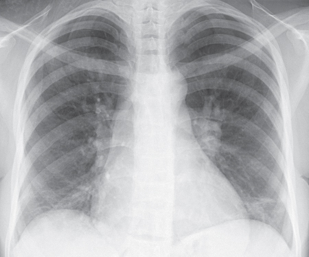
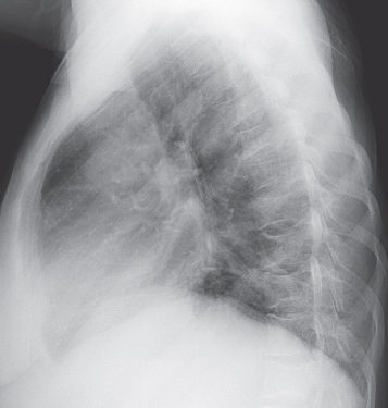
CASE 147 19-year-old man with known sickle cell anemia and new onset of cough, fever, and chest pain PA (Fig. 147.1) and lateral (Fig. 147.2) chest radiographs demonstrate moderate cardiomegaly, low lung volumes, and bilateral basilar linear opacities. Note characteristic H-shaped endplate vertebral deformities in the thoracic spine (Fig. 147.2). Sickle Cell Disease: Acute Chest Syndrome • Bacterial Pneumonia • Pulmonary Embolism/Infarction Sickle cell anemia is a hemolytic anemia that results from the production of abnormal hemoglobin molecules, which deform the red blood cells and impair their transit through vascular channels. Resultant vascular occlusion produces tissue ischemia and infarction. Acute chest syndrome is the second leading cause of hospitalization in patients with sickle cell anemia. Fig. 147.1 Fig. 147.2
 Clinical Presentation
 Radiologic Findings
 Diagnosis
 Differential Diagnosis
 Discussion
Background
Related posts:


Stay updated, free articles. Join our Telegram channel

Full access? Get Clinical Tree


