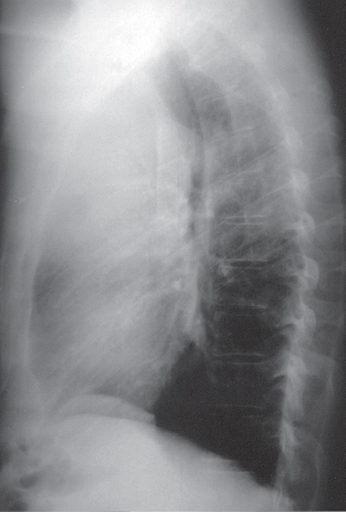
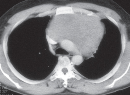
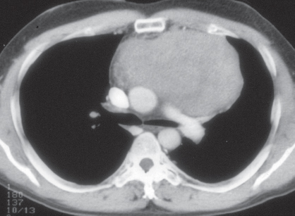
CASE 154 45-year-old man with Cushing syndrome PA (Fig. 154.1) and lateral (Fig. 154.2) chest radiographs demonstrate a large, well-defined anterior mediastinal mass that extends predominantly to the left. Note the significant mass effect on the anterior trachea (Fig. 154.2). Contrast-enhanced chest CT (mediastinal window) (Figs. 154.3, 154.4) demonstrates a large, lobular, slightly heterogeneous anterior mediastinal mass with marked mass effect on the great vessels and the airways. Thymic Carcinoid • Thymoma • Thymic Carcinoma • Lymphoma • Seminoma Fig. 154.1 Fig. 154.2 Fig. 154.3 Fig. 154.4 Thymic carcinoid is a rare primary malignant neuroendocrine thymic neoplasm.
 Clinical Presentation
 Radiologic Findings
 Diagnosis
 Differential Diagnosis

 Discussion
Background
Etiology
Related posts:
![]()
Stay updated, free articles. Join our Telegram channel

Full access? Get Clinical Tree


Radiology Key
Fastest Radiology Insight Engine


