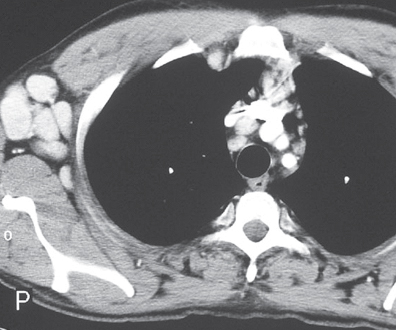
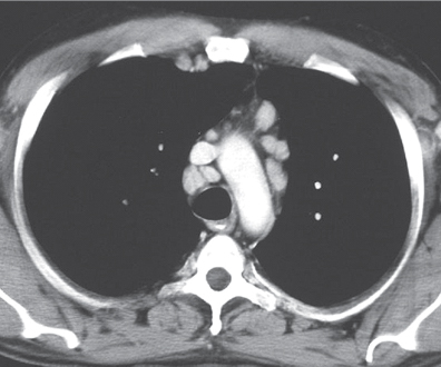
CASE 161 28-year-old man with malaise, fever, night sweats, skin lesions, and palpable supraclavicular lymphadenopathy Contrast-enhanced chest CT (mediastinal window) (Figs. 161.1, 161.2) demonstrates multiple borderline enlarged mediastinal and right internal mammary lymph nodes and enlarged right axillary lymph nodes which exhibit intense contrast enhancement. Multicentric Castleman Disease • Metastatic Disease • Lymphoma • Angioimmunoblastic Lymphadenopathy Fig. 161.1 Fig. 161.2 (Reproduced with permission from Parker MS. Multicentric hyaline-vascular Castleman disease. Clin Radiol 2007; 62:707–710.) Castleman disease, also known as angiofollicular or giant lymph node hyperplasia, is a rare lymphoproliferative disorder characterized by lymph node enlargement with extensive capillary proliferation. Two forms of Castleman disease are recognized, localized or unicentric (the most common) and multicentric or disseminated
 Clinical Presentation
 Radiologic Findings
 Diagnosis
 Differential Diagnosis


 Discussion
Background
![]()
Stay updated, free articles. Join our Telegram channel

Full access? Get Clinical Tree


