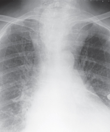
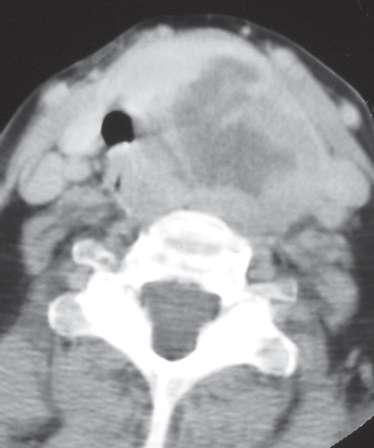
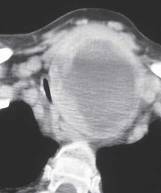
CASE 164 Elderly woman with respiratory distress and a chronic left neck mass Coned-down PA chest radiograph (Fig. 164.1) demonstrates a large, well-defined left-sided mediastinal soft-tissue mass and an associated ipsilateral neck mass. Note marked mass effect on the cervical and intrathoracic portions of the trachea. Contrast-enhanced chest CT (mediastinal window) (Figs. 164.2, 164.3) shows a large, heterogeneously enhancing soft-tissue mass that arises from the left lobe of the thyroid gland (Fig. 164.2) and extends into the mediastinum (Fig. 164.3). Note the large area of central low attenuation surrounded by irregular enhancing soft tissue and significant mass effect on the trachea, esophagus, and mediastinal great vessels. Mediastinal Goiter • Thyroid Carcinoma • Lymphadenopathy; Lymphoma • Neurogenic Neoplasm Fig. 164.1 Fig. 164.2 Fig. 164.3 Mediastinal (intrathoracic, substernal, retrosternal) goiter refers to thyroid tissue within the mediastinum. Mediastinal goiter affects approximately 5% of the world population. The term substernal goiter
 Clinical Presentation
 Radiologic Findings
 Diagnosis
 Differential Diagnosis
 Discussion
Background
Related posts:
![]()
Stay updated, free articles. Join our Telegram channel

Full access? Get Clinical Tree


Radiology Key
Fastest Radiology Insight Engine


