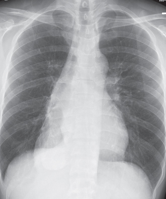
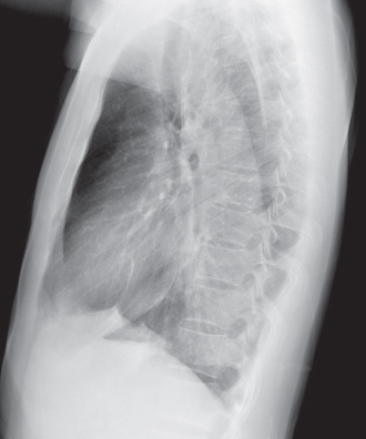
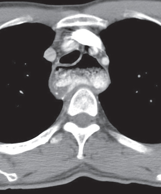
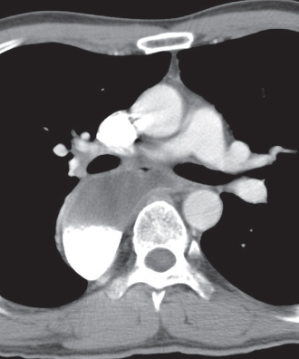
CASE 168 35-year-old man with dysphagia, weight loss, and halitosis PA (Fig. 168.1) and lateral (Fig. 168.2) chest radiographs demonstrate a large, elongate middle-posterior mediastinal mass that extends to the paravertebral region, projects to the right of midline, and produces mass effect on the posterior wall of the trachea (Fig. 168.2). Contrast-enhanced chest CT (mediastinal window) (Figs. 168.3, 168.4) shows heterogeneous contents and dependent contrast in a dilated esophagus that produces mass effect on the heart and tracheobronchial tree (Fig. 168.4). Achalasia • Esophageal Dilatation from Progressive Systemic Sclerosis • Esophageal Dilatation from Acquired Focal Obstruction Fig. 168.1 Fig. 168.2 Fig. 168.3 Fig. 168.4
 Clinical Presentation
 Radiologic Findings
 Diagnosis
 Differential Diagnosis
 Carcinoma
 Metastatic Disease
 Non-Neoplastic Stenosis
Related posts:
![]()
Stay updated, free articles. Join our Telegram channel

Full access? Get Clinical Tree


Radiology Key
Fastest Radiology Insight Engine


