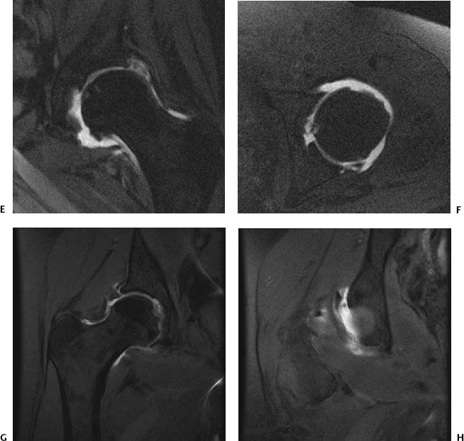
CASE 18 Anthony G. Ryan and Peter L. Munk A patient reported recent onset of hip pain and “clicking” after a forceful twisting injury. Figure 18A Figure 18B Figure 18C Figure 18D T1-weighted coronal oblique (Figs. 18A–18C) and sagittal (Fig. 18D) images after intra-articular injection of dilute gadolinium show linear high signal intensity extending from within the anterosuperior labrum to the surface, present over a series of contiguous slices. Acetabular labral tear. The clinical presentation has a differential diagnosis, including The imaging appearance has no differential diagnosis. The acetabular labrum is a fibrocartilaginous ring applied to the outer acetabular fossa margin, serving to increase hip joint stability by deepening the fossa. The increased depth promotes development of negative hydrostatic pressure within the joint, further increasing the “hold” of the acetabulum on the femoral head. Injuries to this structure therefore have significant effects on the stability of the joint. Although trauma is the unifying cause in all cases, certain conditions predispose to developing early or severe labral tears, including femoral acetabular impingement, congenital/developmental hip dysplasias (Figs. 18E, 18F) and following posterior hip dislocation (Figs. 18G, 18H). In dysplasias, it is thought that tears occur as a degenerative phenomenon secondary to abnormal force redistribution within the joint. The causative trauma is not necessarily severe, often involving a simple twisting or slipping mechanism, such as occurs when one tries to catch one’s balance on ice or a slippery floor, or recurrent low-grade trauma at the extremes of hip flexion. The labrum is well enervated and thus is very painful when injured or chronically irritated. In addition to the physical instability induced by a defect in the labrum and consequent loss of uniform intra-articular pressure, injury to the abundant proprioceptive supply decreases the stability of the joint. The anterosuperior portion of the labrum is torn most frequently. The labrum is most commonly avulsed from the bony acetabulum rather than a true intrasubstance labral tear.
Acetabular Labral Tear
Clinical Presentation
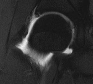
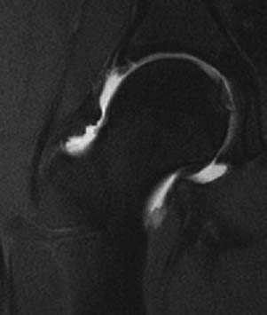
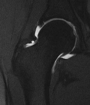
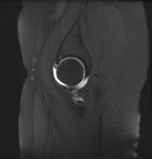
Radiologic Findings
Diagnosis
Differential Diagnosis
Discussion
Background
Etiology
Pathophysiology
Related posts:
Stay updated, free articles. Join our Telegram channel

Full access? Get Clinical Tree


