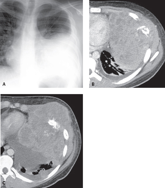
CASE 181 17-year-old man with left chest pain and dyspnea Coned-down PA chest radiograph (Fig. 181.1A) shows a mass-like opacity in the left hemithorax and a left pleural effusion. Contrast-enhanced chest CT (mediastinal window) (Figs. 181.1B, 181.1C) demonstrates a large heterogeneous mass invading the left anterolateral chest wall and destroying the anterolateral aspect of the left sixth rib. The mass contains small areas of indistinct calcification. Note moderate-size left pleural effusion (Figs. 181.1B, 181.1C). Askin Tumor/Primitive Malignant Neuroectodermal Tumor (PNET) Fig. 181.1 • Metastatic Tumor • Lymphoma (direct extension) • Infection (e.g., tuberculosis) Primitive neuroectodermal tumors (PNETs), originally known as Askin tumors, are malignant small round cell tumors that typically affect children and adolescents, but may be encountered in adults. They are typically unilateral, aggressive masses that originate in the chest wall, destroy bone, and are typically associated with pleural effusion. Women are affected more commonly than men (3:1). Detection of associated rib destruction establishes the tumor location within the chest wall. In an adult, the most common etiologies of rib destruction are metastatic lesions, multiple myeloma, and chondrosarcoma. In children and adolescents, the most likely neoplasms include Ewing sarcoma, neuroblastoma, lymphoma, and Askin (PNET) tumor.
 Clinical Presentation
 Radiologic Findings
 Diagnosis

 Differential Diagnosis
 Discussion
Background
Etiology
Stay updated, free articles. Join our Telegram channel

Full access? Get Clinical Tree


