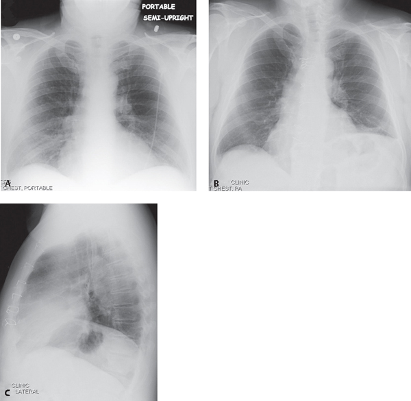
CASE 185 45-year-old man 6-weeks status post CABG with complaints of increasing dyspnea on exertion and orthopnea who cannot tolerate lying flat and must sleep with his head elevated on 4–5 pillows Preoperative frontal chest X-ray (Fig. 185.1A) shows mild cardiomegaly and a normal relationship of the right and left diaphragms. No underlying lung disease is present. Follow-up postoperative frontal (Fig. 185.1B) and lateral (Fig. 185.1C) chest radiographs demonstrate marked elevation of the left diaphragm relative to the right. Note the median sternotomy. Subsequent fluoroscopic “sniff test” revealed paradoxical motion of the left diaphragm. Fig. 185.1 Paralyzed Left Diaphragm • Eventration of the Diaphragm • Elevation of the Diaphragm • Diaphragmatic Hernia (see Cases 94 and 184)
 Clinical Presentation
 Radiologic Findings
 Diagnosis
 Differential Diagnosis
 Congenitally thin muscular portion of diaphragm; appearance increases with age
 5R:1L
5R:1L
 Anteromedial on right; usually involves entire left diaphragm
 Subpulmonic Effusion (see Case 171)
 Atelectasis
 Hypoplastic Lung
 Abdominal Disease (e.g., subphrenic abscess; liver mass; ascites)
 Idiopathic
 Discussion
Background
Related posts:


Stay updated, free articles. Join our Telegram channel

Full access? Get Clinical Tree


