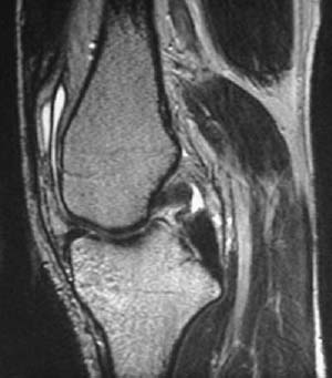
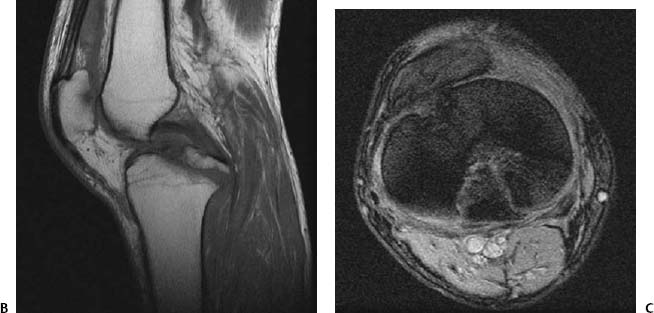
CASE 2 Anthony G. Ryan and Peter L. Munk A 28-year-old man presented with knee pain and swelling after a high-speed skiing injury. Figure 2A A sagittal T2-weighted image of the knee (Fig. 2A) revealed a focal discontinuity of the posterior cruciate ligament (PCL) and a small joint effusion. Acute tear of the PCL None. The PCL stabilizes the knee by preventing anterior translation of the femur on the tibia. It takes its origin from the posterolateral aspect of the medial femoral condyle and attaches on the posterior tibia ~1 cm distal to the articular surface. It is intracapsular but extrasynovial, lying in the same synovial compartment as the anterior cruciate ligament (ACL). The ligament tears most frequently in its midsubstance (50 to 75%). PCL tears occur in isolation in only ~30% of injuries, with 30 to 35% associated with a femoral avulsion and 20 to 30% associated with a tibial avulsion. The most frequent complex of injuries is that of a torn PCL, an associated ACL tear, and a posterolateral corner disruption (including the posterolateral capsule and the popliteus tendon). The usual mechanism of action is that of a direct force on the tibia in a flexed leg, for example, against a dashboard in a sudden-deceleration motor vehicle accident. The ligament is less commonly injured at the extremes of flexion and extension (as is the mechanism in skiing injuries). If a motor vehicle accident is the cause, typically the leg strikes the dashboard, driving the proximal tibia backward, stretching and tearing the PCL. In this setting, bone contusions will be evident at the anterior tibial plateau and the posterior femoral condyles. If there is a hyperextension injury, there is likely to be a tibial attachment avulsion with preservation of the PCL itself (Figs. 2B,2C). The PCL is at particular risk in a posterior dislocation of the knee. Rarely, the PCL may tear in association with closed fractures of the femoral shaft (found in one study to occur in 21% of such fractures), although in this setting, the medial collateral ligament is the most frequent accompanying ligamentous injury (38%). If a direct force on the anterior tibia has been the mechanism, a pretibial hematoma may be present, and there may be an associated fracture of the hip or patella, secondary to the same impact. Figures 2B Midline sagittal T1-weighted image shows a continuous posterior cruciate ligament with an attached avulsed fragment from the tibial insertion. 2C
Posterior Cruciate Ligament Tear
Clinical Presentation

Radiologic Findings
Diagnosis
Differential Diagnosis
Discussion
Background
Etiology
Clinical Findings

![]()
Stay updated, free articles. Join our Telegram channel

Full access? Get Clinical Tree


2 Posterior Cruciate Ligament Tear
