- General cell structure, including specific organelles and their main functions
- The key points of the cell cycle
- Location of cell cycle checkpoints and the role of p53
- Membrane transport and junctions
- Description of mitosis and meiosis
- Cell death
Introduction
The basic unit of living organisms is the cell. Cells were discovered by Robert Hooke in 1665 and described as the functional unit of life. Cells have been called many things, including the smallest unit of life and the building block of life. Unicellular organisms, such as bacteria, contain only one cell throughout their lifespan whereas multicellular organisms, including humans, contain many living cells. The human body contains about 100 trillion, or 1014, cells at any given time.
In 1839, the cell theory was developed by Matthias Jakob Schleiden, Rudolf Virchow, and Theodor Schwann. The cell theory states that organisms are composed of at least one cell, and these cells originate from preexisting cells. In addition to the implication of cell division, the theory states that vital functions within the organism occur in the cell and cells contain hereditary information.
This chapter discusses the basic structure of a cell. Cell structure not only includes construction of the cell itself but also the steps taken by the cell to proliferate.
Cell Structure
Cells are the basic unit of life. All organisms, whether unicellular or multicellular, begin with the function of a single cell. Cells, in general, can be divided into two types: prokaryotic, such as bacteria, and eukaryotic, such mammals. Two features separate these cell types. Prokaryotic cells contain a single external membrane to separate internal and external environments, whereas eukaryotic cells contain internal membranes in addition to their outer membrane. The internal membranes enable the cell to compartmentalize and isolate chemical reactions. In addition to internal membranes, eukaryotic cells have a cytoskeleton. The cytoskeleton provides internal structure and support for the cell. Prokaryotes lack a cytoskeleton.
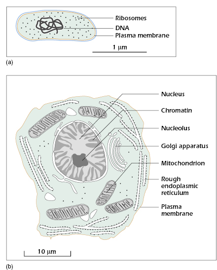
As seen in Fig. 2.1, prokaryotic cells, or bacteria cells, do not contain a nucleus. The DNA is free to move in the cytoplasm. Eukaryotic cells, or mammalian cells, do contain a nucleus. The nucleus is a membrane-enclosed organelle that protects DNA from other organelles in the cytoplasm. Only mammalian cells will be discussed here.
Figure 2.1 (a) Prokaryotic cells (bacterium), shown at the top, do not contain a nucleus. Genetic material is not isolated in the cell but free in the cytoplasm. (b) Eukaryotes (animal cell), on the other hand, isolate DNA within the nucleus, shown at the bottom. From Bolsover et al. (2011), figure 1.9, p. 10.
Mammalian cells are generally split into two cell types. These are the somatic and gamete cells. Somatic cells form tissue and do not participate in reproduction. The tissue is ultimately used to provide life and movement to the organism. Gamete cells are only involved in reproduction. Formation of a gamete cells takes genetic material of the organism and packages it into specialized cells. These cells, when combined, are the beginnings of a new organism.
All mammalian cells contain cytoplasm. Cytoplasm is located between the outer plasma membrane and the nucleus. It is the aqueous phase of cells that contains a wide variety of solutes. The solutes consist of inorganic ions, building blocks of organic constituents, and intermediates of metabolic pathways, to name a few. The cytoplasm can be considered the gas station of the cell. When an organelle needs to refill its supply of a specific ion, that ion is transported from the cytoplasm into the organelle.
Located within the cytoplasm are the organelles of the cell, including the nucleus. The organelles have a wide range of functions from construction of proteins to energy production. Although the number of organelles is great and varies between tissue types, only a select few will be discussed here.
Ribosomes are the protein factory of the cell. The nucleus sends a sequence of messenger RNA (mRNA) to the ribosomes in one of two locations. If the protein sequence is for the endoplasmic reticulum (ER), the molecule travels to the rough ER where bound ribosomes directly insert the protein to the lumen of the ER. However, if the protein is for another purpose, free ribosomes perform protein synthesis in the cytoplasm. In essence, mRNA is the blueprint for protein synthesis, and ribosomes are the general contractors that build from the blueprint.
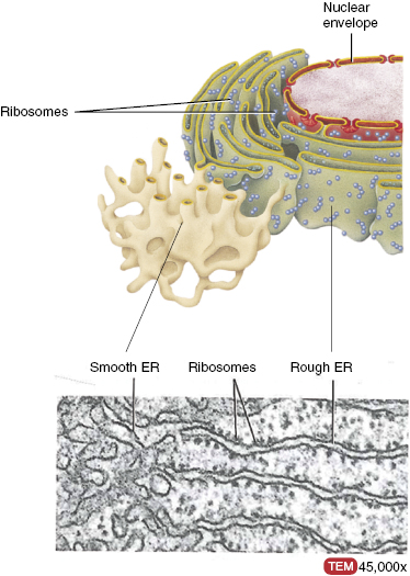
The ER is the first stop for most proteins synthesized in the cell. It is a system of membrane-lined channels that stretch from the nucleus to the cell surface. Within the membrane convolutions, the ER is separated into three distinct regions: the rough ER, the smooth ER, and the transitional region. The rough ER is studded with ribosomes and is involved in the synthesis and folding of proteins. Ribosomes are not found on the smooth ER. It is dedicated to enzyme activity for metabolism of drugs, steroid synthesis, or calcium storage. The differences are shown in Fig. 2.2. Once the rough ER completes protein folding, proteins are transported to the transitional region. In the transitional region, vesicles used for protein transportation are formed. The transitional region is similar to a post office. It takes the folded proteins, packages them according to destination, and sends the package for delivery.
Figure 2.2 The endoplasmic reticulum (ER) is found in two forms: the rough and smooth ER. Rough ER contains embedded ribosomes that assist with protein synthesis, seen on the right, while smooth ER lacks ribosome, seen on the left. The transitional region is not shown. An electron micrograph (bottom) shows a transverse section of the ER. From Tortora and Nielsen (2012), figure 2.10, p. 39.
The abundance of ER varies between each cell type and function. Cells that specialize in the synthesis, storage, and secretion of proteins are historically richer in rough ER. For example, the pancreas is highly specialized in the synthesis and secretion of insulin and glucagon. Pancreatic cells will, on average, contain an area of ER that exceeds the surface area of the outer membrane.
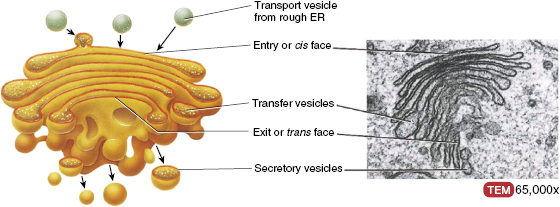
Once the ER has completed protein folding, the protein is secreted to the Golgi apparatus. The Golgi complex is most commonly found within the cell as a stack of three, or more, compartments located near the nucleus. The stack looks similar to a stack of pancakes. Each compartment of the Golgi complex performs a primary function; however, enzymes located in the complex can translocate, which suggests that the compartments are not absolute in function. The Golgi apparatus is responsible for modifying proteins before use by the cell. Some protein modifications are simply the addition or removal of sugar residues and proteolytic processing. Proteolytic processing involves cleaving full-length proteins into smaller, functional components. As seen with the ER, secretory cells have larger Golgi apparatus than nonsecretory cells.
In general, the Golgi apparatus contains a receiving side and a shipping side. The receiving side fuses with transport vesicles from the ER and begins protein modification. Once the modifications are complete, the Golgi complex packages the new protein and sends it into the cytoplasm via the shipping side. The basic structure of the Golgi complex is shown in Fig. 2.3. In addition to modifying proteins made by the ER, the Golgi apparatus synthesizes lipids and cholesterol to be used in plasma membranes.
Figure 2.3 Golgi apparatus is divided into separate compartments that appear as a stack of pancakes, each of which perform specific functions. One side of the Golgi receives the folded protein, the middle section processes the protein, and the opposite side ships the final protein to its destination. The general movement of protein from the ER through the Golgi from the receiving side through the secretory side. An electron micrograph (right) of a transverse section of the Golgi complex shows multiple layers of the organelle. From Tortora and Nielsen (2012), figure 2.11, p. 40.
Lysosomes are the digestive organelle of the cell. The lysosome contains specialized enzymes that break down proteins, DNA, and some carbohydrates. When lysosomes receive extracellular, or intracellular, particles and solutes, hydrolytic enzymes break the particles down. The constituents are transported across the lysosomal membrane to be reused through synthesis of other macromolecules. If the by-products are not needed, they are transported to the outer membrane and released into the extracellular space. Since cells are made of proteins and carbohydrates, the lysosome could potentially digest the cell itself. However, the membrane surrounding the lysosome is constructed to withstand the inner hydrolytic enzymes and prevent degradation of the cell itself.
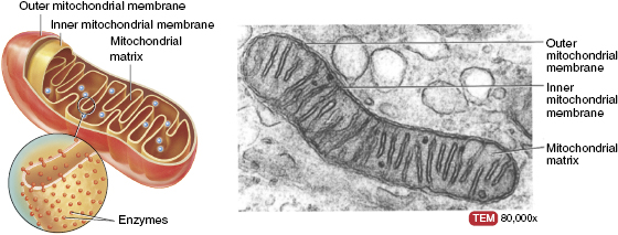
The power house of the cell is localized in the mitochondria. Mitochondria produce energy for all cellular functions through catabolizing, or breaking down, nutrients taken into the cell. The elliptically shaped mitochondria contain a second inner membrane that folds into the central cavity. Specific enzymes necessary for energy production are located, and specifically arranged, within the folds of the inner membrane. Figure 2.4 shows the numerous folds of the mitochondrial inner membrane. The specific enzyme arrangement facilitates an efficient sequence of catabolic reactions needed for energy production. As seen with the ER and Golgi complex, the number of mitochondria per cell greatly depends on the energy requirements of that cell. Cardiac muscle, for example, requires high levels of energy to function properly and many mitochondria are present to fulfill that need. On the other hand, the low energy requirements of lymphocytes can be met with only a few mitochondria.
Figure 2.4 The mitochondria contain two clearly separate membranes. The inner membrane is highly convoluted and responsible for energy production. Enzymes are specifically located within the mitochondrial matrix for efficient production of energy. An electron micrograph (right) of a transverse section of the mitochondria clearly shows the folds of the inner membrane. From Tortora and Nielsen (2012), figure 2.14, p. 43.
Most of the proteins and enzymes found within mitochondria are transported from the cytoplasm. However, mitochondria do contain some DNA and ribosomes to produce proteins. DNA found within the mitochondria contains the sequence for proteins that contribute to the assembly of the organelle itself. Although DNA can be found within the mitochondria, it is not involved in cellular replication. Mitochondrial DNA is solely maternal in origin.
The nucleus is the most complicated of all organelles found within mammalian cells. It is the control center of the cell and contains the genetic material, or DNA, of the cell. Along with DNA, the nucleus contains machinery required for DNA replication and RNA transcription and processing. The nucleus is enveloped in a double membrane to effectively separate the DNA, RNA, and replication machinery. The outer nuclear membrane is fused with the outer membrane of the ER. The inner membrane of the nucleus is used to separate immature RNA from the translation apparatus. Figure 2.5 shows both nuclear membranes along with the connection between the ER and the nucleus.
Figure 2.5 Nuclear envelope showing the nuclear membranes and the connection between the ER and the nucleus. From Tortora and Nielsen (2012), figure 2.15, p. 44.
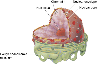
Within the nucleus lies the nucleolus. The nucleolus is responsible for surrounding transcriptionally active regions of RNA, specifically, RNA that contains the sequence used to form ribosomal bodies. Recall from earlier discussion that ribosomes are used to interpret mRNA from the nucleus and synthesize proteins for use in the cell. The nucleolus also processes other stable RNA.
The nucleus houses DNA molecules, which are extremely long and called chromosomes. Chromosomes contain all the genetic material needed for life. Transcription of DNA leads to the production of mRNA. The mRNA then delivers a sequence of nucleotides to the rough ER, and synthesis of a protein begins in the ribosomes.
Located just outside the nucleus are two centrioles. Centrioles are rod-shaped structures that are perpendicular to each other. During cell division, the centrioles migrate to opposing ends of the cell. Centrioles organize spindle fibers that are involved in chromosome separation during cellular division, discussed later.
Cell Membranes
Cellular membranes, or plasma membranes, separate the interior of cells from the external environment. The membrane is selectively permeable, meaning that only certain ions and molecules can enter the cell. Movement into and out of the cell is closely regulated by the plasma membrane. The basic structure of the plasma membrane is a phospholipid bilayer that is embedded with cholesterol and protein. The cholesterol acts as a stabilizer for the membrane by reducing the phospholipid fluidity. Proteins are mostly utilized for cellular transport and junction formation. The fluid mosaic model is the best model for plasma membranes.
In 1972, S.J. Singer and Garth Nicolson came up with the idea of treating plasma membranes as two-dimensional entities. In their model, lipid and protein molecules are able to diffuse more or less easily across the membrane. In Fig. 2.6, the fluid mosaic model of a cellular membrane is shown. What has been discovered since 1972 are three basic domains within the membrane, each with a specific function. These are protein–protein complexes, lipid rafts, and pickets and fences.
Figure 2.6 A typical plasma membrane demonstrating the phospholipid bilayer, with associated cholesterol and protein complexes. From Tortora and Nielsen (2012), figure 2.2, p. 30.
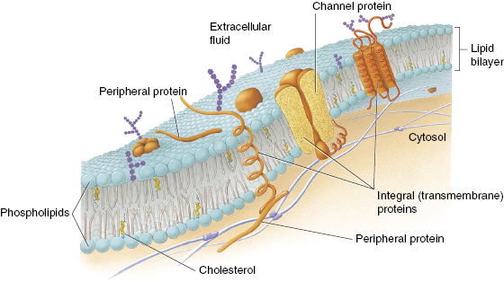
Protein–protein complexes are used mainly for transportation across the plasma membrane. The protein complexes can be located across the membrane, on one side of the bilayer or peripheral. These complexes act as ion channels, proton pumps, or cellular receptors. Lipid rafts are tightly packed, highly organized, groups of lipids that float freely within the plasma membrane. The lipid rafts serve as organization centers for molecule assembly and protein trafficking, among other duties. Pickets and fences, the last domain, are the scaffolding of the cell. They are made of cytoskeleton proteins and maintain the overall structure and shape of the cell.
In addition to the outer plasma membrane, each organelle is surrounded by a membrane. Organelle membranes separate the functional cavities of the organelle from the cytoplasm of the cell. The composition of each organelle membrane depends on the function of the organelle. Lysosomes, as mentioned previously, are responsible for breaking down proteins and carbohydrates. The acidic nature of lysosomal enzymes requires a specialized membrane to prevent leakage into the cytoplasm.
The most specialized membrane is the nuclear envelope. The nuclear envelope is a selectively permeable membrane that separates the nucleus from the cytoplasm. Nuclear pores fuse the two membranes together. These pores control movement of macromolecules into and out of the nucleus. One purpose of the nuclear pores is to ensure that only fully processed mRNA is delivered to ribosomes for protein synthesis.
Related posts:
Stay updated, free articles. Join our Telegram channel

Full access? Get Clinical Tree


