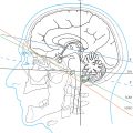
2 Tomography and Landmarks
CT and MRI are widely used cross-sectional imaging modalities offering excellent imaging capabilities for the head and spine.
2.1 Computed Tomography
The spiral (helical) or volume technique is employed by modern CT devices. Secondary slices in a required 3D orientation and of desired thickness can be reconstructed from data obtained from transverse scanning of the examined measuring volume.258 , 272 , 282 , 344 , 350
The image is displayed following the measurement of the X-ray absorption of voxels in the examined volume. The X-ray density of tissue of interest is specified in HU. This is assigned in accordance with certain default values.
The so-called partial volume effects may be minimized by a high-resolution matrix and minimal slice thickness. Partial volume effects, image artifacts, and prevailing examination conditions (contrast medium administration, restlessness, positioning) result in distinctive features that must be kept in mind during image evaluation.258 , 272 , 396 , 410 , 476 , 583 Artifacts may be reduced by setting the imaging plane while avoiding beam hardening, as well as by the addition of several thin slices to a slice in the desired thickness.258
Intravenous (IV) administration of iodinated contrast medium improves the informative value of the examination by opacifying both physiological (blood vessels) and pathological structures (many tumors, inflammation, etc.).250 , 476 A CTA may be performed using IV contrast medium administration as bolus with faster CT devices (short sampling times, continuous helical or volume measurement) and a shorter examination time. Smaller vessels of the brain may thus be depicted by data acquisition in spiral technique either as so-called 3D surface reconstruction, by MIP or by volume rendering processes.258 , 272 , 275 , 499 , 583
2.2 Magnetic Resonance Imaging
The magnetically effective angular momentum (spin) of atomic nuclei with an odd number of nucleons (protons and neutrons) is used to generate MR images. The hydrogen nucleus possesses a large magnetic moment and is frequently present in organisms. Thus water-containing tissues as well as lipids and proteins with their high hydrogen atom content are particularly well visualized on MRI.134 , 272 , 350 , 364 , 441 , 557 , 583 , 657
For a given magnetic field strength, the MR signal is determined by proton density, T1 relaxation time (spin lattice or longitudinal relaxation time), T2 relaxation time (spin–spin or transverse relaxation time), and by proton movements in the measuring volume. Signal intensity of each measuring volume (voxel) determines the gray-scale value of a pixel on the monitor.
Various sequences may be employed to induce excitation of hydrogen nuclei. These have an effect on image contrast and thus significantly influence the diagnostic value. The SE technique found wide acceptance within the first years since its inception. T1-weighted images and sequences enable exceptional depiction of anatomical details due to a favorable signal-to-noise ratio. CSF has a low T1 signal and is therefore dark, while it exhibits a high signal on T2 images and is consequently bright. Many pathological changes are well seen on T2-weighted images. T1 and T2 sequences are part of the basic configuration of MRI equipment. These have been selected as reference sequences in this book due to their wide availability and good contrast between gray and white matter.
Gradient echo sequences (Flash, FISP, etc.) enable shortening of examination times and reduction of movement artifacts while enabling depiction of flow and tissue perfusion with high temporal resolution and mapping of vessels (MRA).
The diagnostic problem to be addressed determines the selection of measurement criteria of an MRI examination. These include the volume to be examined, slice position, and measurement parameters, for example, slice thickness and interval, matrix, and the sequence to be employed. Critical picture elements must be well delineated and free of artifacts (so-called quality criteria). The cranium should be examined using T1- and T2-weighted sequences, thereby gaining high-contrast imaging of gray and white matter. Additional T2*-weighted sequences or those with greater susceptibility weighting (e.g., SWI) are indicated in certain situations, for example, in cerebrovascular disorders.287 The possibility of potential errors due to artifacts and inadequate performance of the examination is significantly greater than with other imaging techniques due to the large number of modifiable and interdependent measuring parameters. It is therefore not only technical quality assurance, but also one’s medical knowledge which plays a vital role in deciding indication, implementation, evaluation, and assessment of MRI examinations.273 , 563
Several parameters (proton density, relaxation times, etc.) of examined tissue are used in MRI for image acquisition.156 , 516 , 557 , 657 Image processing and reconstruction takes place in MRI in a similar fashion to CT, with the examiner selecting the window center (center) and window width (window). This determines the informative value of the examination, either contributing to image interpretation or taking away from it.
Paramagnetic substances (e.g., gadolinium-DTPA, gadolinium-diamide, manganese, and others) when used as contrast medium improve clinical information in certain cases. They shorten the T1 relaxation time in their areas of distribution, which in turn results in an increase in lesional signal intensity on T1 images as compared to their surroundings.265 , 439 , 557 , 655 A noncontrast T1 examination should be carried out prior to the contrast injection using the same settings.240 Contrast medium administration in general provides the following information:134
Presence or absence of contrast enhancement of a lesion
Extent of the area exhibiting contrast enhancement
Enhancement pattern of this region
Increase or decrease in lesional perfusion using dynamic examination
Structures of the skull base and the infratentorial region as well as the spinal canal are better appreciated on MRI than on CT due to higher soft tissue contrast and lack of interference by bony artifacts. That MRI has been rapidly accepted is justified by its high sensitivity for pathological cerebral and spinal processes due to its exceptionally good contrast resolution.557 MRI is well suited for the depiction of myelination of the central nervous system in young children and may therefore be used in the evaluation of disorders of myelination, myelogenesis as well as diffuse or focal demyelination.30 , 31 , 211 , 272 , 296 , 441 , 558 , 590 MRI in combination with its morphological and functional data is almost equal to CT and CTA in the differential diagnosis of acute stroke and evidence of hemorrhage, but superior to these modalities in excluding tumors. An infarcted area may be demonstrated as early as a few minutes after vascular occlusion using a diffusion-weighted sequence.
In vivo MRS is not yet in common use due to the complex technology involved,475 , 644 , 658 even though several promising observations have been made.126 , 330 , 364 In vivo MRS has a high specificity for the tissue in the measuring volume due to detection of the so-called chemical shift of molecules in the magnetic field. Not only is it suited for the identification of abnormal tissue (tumors, inflammation, necrosis, etc.) in the central nervous system, but also contributes to tumor grading.230 , 232 , 330 , 331 , 419 , 420
fMRI has played an important role in the neurosciences for years.78 , 129 , 214 , 294 , 398 , 425 It helps mapping local changes in cerebral perfusion due to vasoneuronal coupling following targeted motor, visual, or sensory stimulation and may be used to localize short-term changes in brain function under defined paradigms.272 , 563 The technique is time consuming and the signal yield is low, so clinical use so far has been restricted to some very stable paradigms (motor, visual, auditory, and tactile paradigms; speech comprehension and production). Increasingly higher cognitive functions may be understood using higher field strengths and other methods including resting-state fMRI (see ▶Section 10.13).25 , 58 , 118 , 207 fMRI can, for instance, guide a decision for or against cochlear implantation, and also aid in selecting the side for this procedure.528
Related posts:


Stay updated, free articles. Join our Telegram channel

Full access? Get Clinical Tree


