1 Introduction
1.1 Aims and Objectives
Computed Tomography and MRI allow rapid and detailed diagnosis of circulatory disturbances, intracranial bleeding, other space occupying lesions as well as disorders of CSF circulation. These imaging methods are therefore of prime impor tance in the diagnostic armamentarium.
Modern neuroimaging is moreover employed for objective follow-up, for example, in documenting spontaneous regression of subdural hematomas and hygromas or the need for surgery thereof. MRI is an invaluable clinical tool in the diagnosis and treatment follow-up of multiple sclerosis and has furthered the development of new therapeutic concepts and monitoring of their efficiency. MRI being highly sensitive allows detection of small, millimeter-sized tumors (e.g., pituitary microadenomas, intracanalicular vestibular schwannomas) so that these may be successfully removed by microsurgery without loss of function.
Alongside diagnostic radiology, interventional neuroradiology has also gained in importance. This allows generally safer treatment of acute circulatory disorders in strokes and vascular malformations like aneurysms and arteriovenous fistulae, as compared to open surgery.
Widespread availability of the fascinating diagnostic capabilities of modern imaging methods may actually prove disadvantageous and even harmful for patients. It is not always possible to diagnose or exclude all intracranial, cerebral, or meningeal diseases with CT or MR. Unilateral symptoms do not always have a cerebral origin. Pathological findings evident on imaging at first sight are not always responsible for the patient’s symptoms. Correlation between pathological findings seen on imaging and the patient’s symptoms requires functional and topical knowledge of anatomy, which the present form of this book aims to facilitate.
The acquisition of knowledge in neuroradiology, neurology, neurosurgery, psychology, psychiatry, and neuroscience is an on-going process. Pathological findings have neither been described nor depicted in our illustrations, for which one would need to refer to relevant neuroradiological textbooks.22 , 87 , 258 , 272 , 441 , 511 , 557 , 653 , 654 , 655 Every referring physician and radiologist should be familiar both with merits and with limitations of neuroradiological techniques. This would prevent wrong investigations being performed at the wrong time and at the wrong site. Both the examiner and the referring physician should be aware of concrete facts pertaining to pathogenesis of disease (age of hemorrhage or an infarct, also inflammation, etc.).
The neurosciences continue to have high expectations of neuroradiology and further advancements therein; this applies especially to MRS, further developments in spiral CT and particularly to MRI. We therefore placed MR and CT images of nearly the same size as large-format anatomical atlas illustrations side-by-side. Some atlas images required correction of details in instances where new or more precise information was available in literature or fresh insight was obtained through our own studies.
Tomographic images of the human body are obtained using modern digital imaging methods. Depicted slices are usually 1 to 10 mm in thickness. These slices are composed of small cuboids (so-called voxels; the term voxel is a contraction of two words, “volume” and “element”). Its height corresponds to slice thickness and the length of its edge to the image matrix. X-ray absorption and signal intensity are determined for every voxel in CT and MRI, respectively (see Sections 2.1 and 2.2). The calculated measurement from a voxel is assigned a gray value or a color as pixel on a monitor or film base (the term pixel is a contraction of two words, “picture” and “element”).
Transverse (axial) sections (perpendicular to the body’s long axis) are normally examined in CT due to the requisite rotational movement of the scanner. Frontal (coronal) sections are obtained by reformation. One of the advantages of MRI is that the sectional plane may be freely selected. Modern, spiral (helical) CT scanners do not obtain data by scanning objects slice after slice and then processing it. Instead a powerful X-ray tube rotates constantly around the patient’s body while the patient continuously moves through the scanning field. A data set is thus obtained from a large measuring volume (volume scan) and is then secondarily processed to acquire an image. Thus, any slice position and slice thickness may be computed and selected from the volume data set. Scan times of multislice spiral CT lie well under a minute. 3D reconstruction of interesting structures may be obtained using sophisticated software and a powerful workstation. Multiplanar and 3D reconstruction thus allows high resolution visualization of cranial blood vessels (CTA) after IV bolus injection of contrast medium.
Digital cross-sectional imaging methods required reorientation of the depiction of brain anatomy with development of a modified topographical orientation for clinical applications. Traditional neuroanatomy is based on the development of the mammalian brain. The human forebrain developed an obtuse angle with the brainstem from an originally stretched out horizontal position as a result of pronounced brain development during evolution and the upright posture. Neuroanatomy of the human brain is depicted in at least two series of slices in many textbooks: One series of images (so-called frontal series) is aligned perpendicular to the long axis of the forebrain (Forel axis), while the other is perpendicular to the long axis of the brainstem (Meynert axis). This approach is especially practical for comparative neuroanatomy, allowing for a better comparison of neuroanatomical findings in animals with those in humans. The axes of the forebrain and the brainstem in most mammals approach a nearly straight line. In contrast however the two long axes in the human brain form an obtuse angle of about 110 to 120° with each other. There exists, therefore, no ideal reference plane for transverse sections in humans, such that images obtained simultaneously for the forebrain and the brainstem conform to conventional neuroanatomical images.
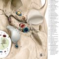
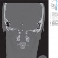
Transverse sections parallel to the bicommissural line are obtained in MRI; this line runs through the midpoint of the anterior and posterior commissures. CT sections are conventionally obtained in a plane tangential to the roof of orbit (supraorbito-meatal line) due to its good reproducibility while allowing protection of the radio-sensitive lens of the eye. In everyday work however the supraorbito-suboccipital line (see ▶Fig. 1.1) is identified more quickly on the scout image (scout, localizer, topogram). This does not differ significantly from the supraorbito-meatal line and helps avoid planning delays resulting from poorly delineated contours of the internal auditory canal due to well pneumatized mastoids.

Ambrose13 preferred the bicommissural plane as the reference plane for CT, which connects the lateral angle of the eyelid (canthus) with the external auditory canal. This historical sectioning plane has not been taken into account in the present new edition. The characteristics of these transverse imaging planes are described in Section 1.2.2.
Obtained intravital images oriented along sections running parallel to the frontal, bicommissural (MRI), and supraorbito-suboccipital planes differ from conventional neuroanatomical images acquired postmortem (see Section 1.3). The benefits of new cross-sectional imaging methods can however be fully exploited only when sufficient knowledge of 3D structures is available. A series of works were thus published in the last three decades, which reproduced macroscopic anatomy in transverse and multiplanar planes.60 , 208 , 219 , 333 , 442 , 532 , 572 Neurofunctional systems cannot however be depicted solely macroscopically since they are composed of macroscopic and microscopic findings and are especially developed from hodological outcomes. Hodology is the study of neuronal interconnections in the central nervous system. Thus, even the most refined techniques of macro- and micro-photography of brain sections preclude the imaging of synaptic circuitry of individual neurofunctional systems.
This book therefore aims at the following:
Depiction of brain anatomy in the atlas section of the book in the three conventional planes used in cross-sectional imaging: Anatomical sections were employed to achieve this end. A drawing technique was developed for quick yet precise orientation, whereby anatomic structures were adapted to gray-scale CT and MR images. Gray scale T1-weighted MR images (see Section 2.2) correspond only partially to graphic illustrations. The illustrations however do not make an allowance for the wide spectrum of MR signal intensities of biological structures, which in turn depend on the selected examination sequence. Corresponding large-format CT and MR images in the atlas section (colored stripes at the edges) should enable readers to draw comparisons. All illustrations in the atlas section were derived from original preparations, see Chapter 12 (coronal and sagittal series, brainstem series), from test subjects (axial MRI series) or patient images (axial CT series). Macrophotography can only reproduce the surface of the slice. In contrast, CT and MRI display the contents of a slice since they transform voxels into pixels, as described. Illustrations are advantageous since important neuroanatomic structures like tracts and nuclei, not visible at the surface since they lie within the slice, may be graphically represented by interrupted lines or hatched areas. Microscopically recognizable structures, such as certain cortical areas, which could not have been depicted in isolation using macrophotography, have also been incorporated in our illustrations. The atlas images of this book (see ▶Fig. 3.2 to ▶Fig. 3.15, ▶Fig. 4.2 to ▶Fig. 4.7, and ▶Fig. 5.2 to ▶Fig. 5.30) were reproduced true to scale in definite coordinate systems in accordance with similar principles such as those in stereotactic atlases6 , 16 , 63 , 514 , 573 , 574 so that anatomical and neuroanatomical structures of the “model brain” could be rendered over a patient’s brain using a definite coordinate system. A detailed explanation for the above is outlined in Section 1.2.
Graphic rendition of principal arterial territories in the supra- and infratentorial regions: Blood vessels and their vascular territories have been depicted in detail in the coronal, sagittal, and transverse planes and compared with angiographic images. This should simplify the interpretation of CTA and MRA images and correlation of CT and MR findings with angiographic images (DSA) for clinicians. A depiction of the variability of vascular territories, which may be enabled by in vivo MRI methods of arterial spin labeling, have deliberately not been touched upon.
Description and imaging of principal neurofunctional systems in multiplanar parallel slices: The main tracts of the neurofunctional systems have been graphically represented in transverse sections, the most important of these additionally in coronal and/or sagittal sections. Equidistant slices were employed for this purpose in a right-angled coordinate system. The distance between two planes of the coronal, sagittal, and bicommissural series was 1 cm and 5 mm in the brainstem series, respectively. True-to-scale illustrations of slices have been positioned in a gray environment in such a manner that they are viewed vertically from below on the bicommissural (MRI) and supraorbito-suboccipital (CT) oriented parallel sections as well as from the front on the coronal and from the left on sagittal parallel sections. The observer can thus imagine a 3D reconstruction of individual structures through true-to-scale reproductions and optimal positioning of individual slices in a 3D coordinate system, as in the drawings in Chapters 6 to 9, or neurofunctional systems, as in the drawings in Chapter 10. A computerized graphic rendition of neurofunctional systems and the large arteries of the brain over and above the selected tomographic images (pertains to the third edition) has been published as a book and as CD ROM312 , 313 where these systems have been rendered in pseudo-3D and 3D. Real 3D reconstructions in different projections and combinations may be acquired on a PC using red–blue glasses.
Elucidation of the relationship of the topographyof neurofunctional systems with the site of a lesion and its clinical symptoms: Individual neurofunctional systems and their cardinal symptoms have been described. This spatial knowledge will define investigations more precisely. Consensus between the clinical syndrome and the lesion detected on tomography confirms the topical diagnosis. Discrepancy between clinical symptoms and CT or MR findings requires a reevaluation of clinical findings and further diagnostic procedures. Moreover, the spatial relationship of the site of the lesion to neurofunctional systems may be indicative of prognostic outcome.
Magnified rendition of the brain stem supplemented by T1w and T2w MEDIC–MR images: Nuclei and tracts lie closely packed together in the brain stem and therefore require magnification for their graphic representation. Thus, a brain stem series in 5 mm slices was appended to the bicommissural oriented axial series in a coordinate system defined by the Meynert plane. Bony artifacts in CT preclude satisfactory imaging of these fine structures. Only with MRI is low-noise imaging of the brain stem possible; signal loss induced by the clivus or the sphenoid sinus may occur in the MEDIC sequence.
Anatomical structures and their variants, relevant for everyday practice, have been depicted in the text of this book. We restricted ourselves to presently available and reliable findings pertaining to neurofunctional systems. Speculative information and not generally accepted research findings have not been taken into account.
The atlas, text, and supplemental illustrations focus on the depiction and description of neuroanatomy of the brain and its neurofunctional systems. The normal topography of the head and the craniocervical junction has also been discussed and depicted in illustrations, as diseases of these regions may spread to the brain and vice versa. Of prime importance is normal cross-sectional anatomy with presently feasible CT and MR representation of intravital anatomical relationships, while keeping functional aspects in mind. Special settings, magnified images or contrast medium administration for optimal imaging of individual regions, or structures (pituitary, blood vessels, etc.) have been dispensed with. Graphic representation and rendition of MR and CT images of pathological changes have been intentionally omitted. The text provides some information pertaining to pathological changes with clinical correlation. The physician thereby receives suggestions for optimal investigations in order to improve the meaningfulness of the chosen diagnostic study. A suitable imaging procedure may be selected while taking into account examination findings and neurophysiological data, simplifying investigations with a targeted approach and thereby optimizing treatment.
Interdisciplinary knowledge is an important prerequisite for optimal use of and comprehensive assessment by modern cross-sectional imaging methods. While the neuroradiologist requires a functional knowledge of anatomy to be able to deal with clinical problems in a competent fashion, the neurologist, neurosurgeon, radiation therapist, physician, pediatrician, etc. need to be familiar with both capabilities and diagnostic strengths of modern cross-sectional imaging methods. The objective of this book is to bridge the gap between the two in a simplified, universally understood manner.
Related posts:


Stay updated, free articles. Join our Telegram channel

Full access? Get Clinical Tree


