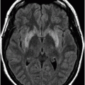
Axial contrast-enhanced CT scan through the level of the ventricles.
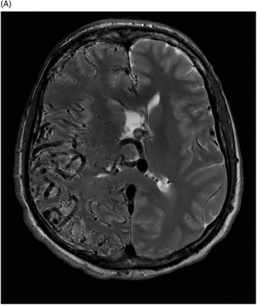
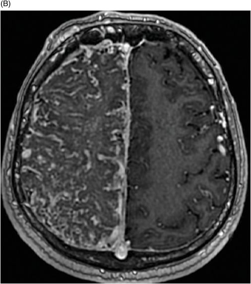
(A) Axial T2WI through the level of the ventricles. (B) Postcontrast axial gradient echo T1WI sequence through a slightly superior aspect of the brain.
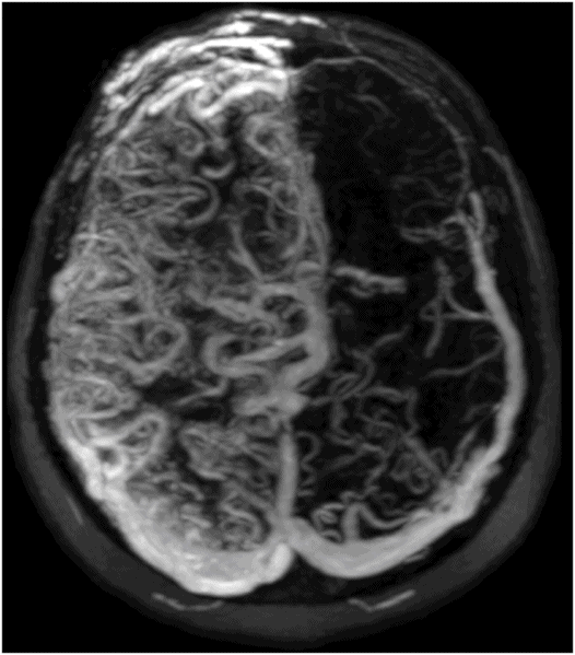
Maximum intensity axial projection of the 3D postcontrast axial T1WI of the head.
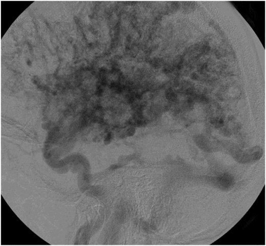
Lateral digital subtraction angiogram of the head.
Cerebral Proliferative Angiopathy
Primary Diagnosis
Cerebral proliferative angiopathy
Differential Diagnosis
Arteriovenous malformation
Imaging Findings
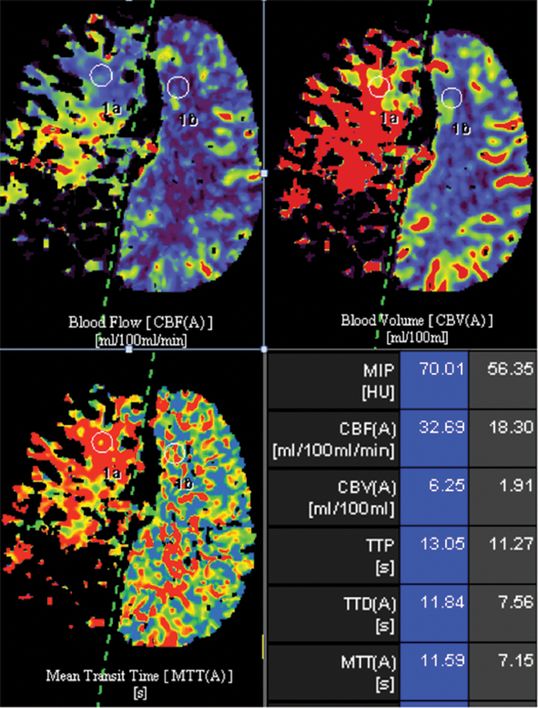
Fig. 21.1: Axial contrast-enhanced CT scan through the level of the ventricles demonstrated numerous, abnormal-appearing blood vessels involving almost the entire right hemisphere with relative sparing of the medial right frontal lobe. Diffuse swelling of the hemisphere, with subtle subfalcine herniation, was also noted. Fig. 21.2: (A) Axial T2WI through the same level demonstrated flow voids from abnormally large blood vessels in the right hemisphere and adjacent to the septum of the ventricles. The intervening brain has a relatively normal appearance. The left hemisphere was normal. (B) Postcontrast, axial gradient echo T1WI sequence through a slightly superior aspect of the brain demonstrated similar findings. Vessels are numerous along the posterior aspect of the brain close to the dura, as well as along the falx, suggesting dural involvement. Abnormal vasculature was also noted in the right frontal scalp. Fig. 21.3: Maximum intensity axial projection of the 3D postcontrast, axial T1WI of the head demonstrated an extensive network of abnormally large blood vessels in the right side of the cranial cavity and in the right frontal scalp. A large vein was also noted along the convexity of the left hemisphere. Fig. 21.4: Perfusion parametric maps with region of interest through the same level as Fig. 21.2B demonstrated increased CBV, CBF, and mean transit time (MTT) in the relatively normal-appearing area of the right frontal lobe. Please note that there was absence of signal over the areas of abnormally enlarged blood vessels on CBV, CBF, and MTT maps (values in these areas were outside the scale showing these results). Fig. 21.5: Lateral digital subtraction angiogram of the head demonstrated a network of abnormally large blood vessels supplied by the right internal carotid artery. The morphology is bizarre: no discrete nidus, feeding artery, or prominent draining vein was noted.
Stay updated, free articles. Join our Telegram channel

Full access? Get Clinical Tree


