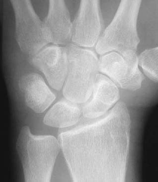
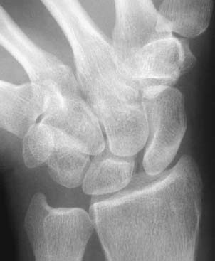
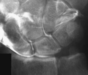
CASE 24 Anthony G. Ryan and Peter L. Munk A 30-year-old man presented to us with complaints of chronic wrist pain and weakness since a motorcycle accident 18 months previously. Figure 24A Figure 24B Figure 24C An AP radiograph with the wrist in the neutral position (Fig. 24A) appears unremarkable initially; however, the scaphoid is foreshortened, and the distal pole appears more prominent than usual, projected as a ring through the distal body of the scaphoid. The arcs of the wrist (arcs of Gilula) are intact. An AP radiograph with the wrist in ulnar deviation shows an increase in the scapholunate (SL) interval (Fig. 24B). An exposure from an arthrogram (Fig. 24C) shows contrast insinuating into the SL interval. Scapholunate ligament tear with rotary subluxation of the scaphoid. Normal variant of ligamentous anatomy, that is, congenital absence of the SL ligament. The SL ligament is typically composed of a dense fibrous volar trapezoidal component; a thin, filmy middle triangular component; and a dorsal component, composed of a dense fibrous band. The dense volar and dorsal components are attached directly to bone and provide the majority of the biomechanical stability of the joint, whereas the thinner middle portion attaches to hyaline cartilage. The SL ligament may be congenitally absent. Through acute trauma, chronic repetitive trauma, or wear and tear, the ligament may become stretched and elongated or perforated. Degenerative perforations tend to involve the thinner central section and are thus rarely mechanically significant. Trauma or degeneration, the latter most commonly secondary to rheumatoid arthritis or calcium pyrophosphate deposition disease. The ligament may stretch and tear in isolation or occur in combination with a scaphoid fracture. In combination with the lunatotriquetral ligament, the SL ligament is responsible for the mechanical and rotational stability of the proximal carpal row. The disassociation of the scaphoid and lunate thus results in altered biomechanics and disrupts carpal stability. SL instability occurs secondary to a tear of the ligament and is frequently associated with a scaphoid fracture. Carpal instability may be either static or dynamic, the latter producing abnormal intercarpal motion. SL ligament tears are almost unheard of in children. Rotary subluxation of the scaphoid may occur after a fall on the outstretched hand, whereby subsequently, on dorsiflexion of the wrist, the scaphoid cannot maintain its normal relationship with the radius. For SL dissociation to occur, the dorsal SL ligament must be torn in addition to a tear of the interosseous SL ligament. In dorsal intercalated segment instability (DISI), the lunate undergoes palmar translation and dorsal angulation, the capitate is dorsally subluxed, and the scaphoid moves in a volar direction. In volar intercalated segment instability (VISI), the scaphoid and lunate are palmar-flexed with a decreased SL angle (< 30 degrees), a normal or increased capitolunate angle (> 30 degrees), and palmar subluxation of the capitate. VISI is usually associated with lunatotriquetral instability, rather than with SL instability.
Intrinsic Ligament Tear (Scapholunate Ligament Tear)
Clinical Presentation
Radiologic Findings
Diagnosis
Differential Diagnosis
Discussion
Background
Etiology
Pathophysiology
Clinical Findings
Related posts:
Stay updated, free articles. Join our Telegram channel

Full access? Get Clinical Tree


