25 Peritoneum and Mesentery


10.1055/b-0034-75818

25 Peritoneum and Mesentery

Herzog\, Christopher

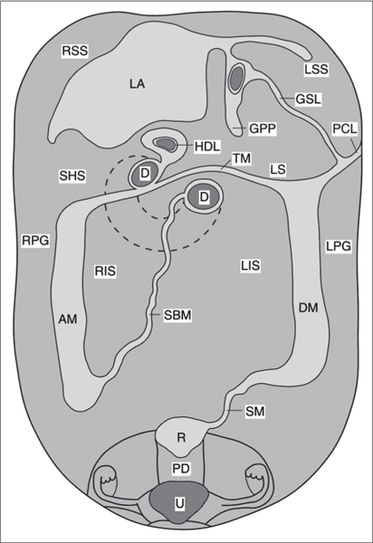
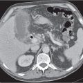
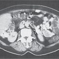
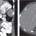
The peritoneal space is an anatomically complex cavity and under normal physiological conditions nonvisible on computed tomography (CT) scans. However, Fig 25.1 displays CT scans of a patient with heavy ascites, which has been made even more attenuating through intraperitoneal contrast application. In combination with Fig. 25.2 , it thus allows for an identification of all relevant peritoneal structures and spaces.


The peritoneal cavity is subdivided by peritoneal plicas into several different compartments and recesses (Fig. 25.2), which are anatomically interconnected.


The transverse mesocolon, which affixes the transverse colon to the retroperitoneum, is a major barrier dividing the abdominal cavity into supramesocolic and inframesocolic compartments. The inframesocolic compartment or space is further subdivided by the small bowel mesentery into the right and left inframesocolic space.


The right inframesocolic space is laterally defined by the peritoneal attachment of the ascending colon, as well as medially and caudally by the mesenteric root.


The left inframesocolic space is laterally and caudally to the left delineated by the peritoneal attachments of the descending colon and the sigmoid. On the right side, it opens caudally into the right paracolic gutter, which lies lateral to the mesenteric attachment of the ascending colon. The left paracolic gutter lies lateral to the peritoneal attachments of the descending colon and the sigmoid. These four inframesocolic spaces and barriers are better visualized on cross-sectional CT scans than the transverse mesocolon.


The supramesocolic space is subdivided into the right and left subphrenic spaces, the subhepatic space, the Morison pouch, and the lesser sac. On the right side, the right paracolic gutter extends cranially into the posterior subhepatic space and the Morison pouch, which represents the most dependent portion of the peritoneal cavity in the right upper quadrant. It also is a frequent site of intraperitoneal fluid collections, which may extend even to the right subphrenic space (supramesocolic space) if the fluid volume suffices. Both paracolic gutters connect the inframesocolic space with the pelvis, where the pouch of Douglas and two lateral paravesical recesses are the most dependent parts and thus frequent sites of intraperitoneal fluid collections.


Because the left coronary ligament lies more anteriorly as compared with the right, the left subphrenic space extends less posteriorly than the right. The lesser sac lies dorsally to the lesser omentum, the stomach, the duodenum, and the gastrocolic ligament but ventrally to the body of the pancreas and the spleen. It is connected with the peritoneal space through the foramen of Winslow, or foramen epiploicum—which hides behind and below the porta hepatis. The lesser sac may extend downward between the anterior and posterior plicas of the greater omentum. The superior recess of the lesser sac usually extends upward to the diaphragm surrounding the caudate lobe of the liver (Fig. 25.1a).


The mesentery of both the small and large bowel and various ligaments provide a barrier not only for the free movement of fluid, cells, and germs but also for the direct spread of diseases within the abdominal cavity. However, in the absence of ascites, identification of individual ligaments may be difficult, as mesentery and intraperitoneal fatty tissue show similar Hounsfield unit (HU) values. Mesentery and ligaments contain blood vessels and therefore may be better identified on postcontrast CT-scans. Mesenteric lymph nodes usually are not discernible due to their predominant fat content. Only reactive nodes appear as well-defined oval or round soft tissue densities.


Nevertheless, several ligaments of the supramesocolic cavity usually can be identified on CT scans.


The right triangular ligament forms from the coalescence of the superior and inferior plica of the right coronary ligament and separates the right subphrenic space from the Morison pouch. The left triangular ligament forms from the superior and inferior plica of the left coronary ligament and is located along the superior aspect of the left hepatic lobe.


The gastrophrenic ligament runs from the dome of the left diaphragm to the stomach and is easily missed on CT.


The gastropancreatic plica (Fig. 25.1b) forms around the proximal left gastric artery as it courses superiorly. It attaches the gastric fundus to the retroperitoneum and partially separates the superior recess of the lesser sac from the splenic recess.


The falciform ligament (Fig. 25.1c) is a remnant of the ventral mesogastrium, which contains the ligamentum teres and the obliterated umbilical vein. This vein may be recanalized in situations with decreased venous hepatic outflow.


The gastrosplenic ligament (Fig. 25.1d) is a remnant of the dorsal mesogastrium, connecting the greater curvature of the stomach with the splenic hilum. It contains short gastric arteries (rami gastrici breves), forms the lateral border of the lesser sac, and may be affected by processes of the stomach or pancreatic tail.


The phrenicocolic ligament is fixed to the spleen and attaches the proximal part of the descending colon to the left hemidiaphragm. By separating the left subphrenic space from the remainder of the peritoneal cavity, it inhibits free flow from the left paracolic gutter to the left subphrenic space. Pancreatic processes can spread via this ligament and involve the splenic flexure of the colon.


The gastrocolic ligament connects the greater curvature of the stomach with the superior aspect of the transverse colon. It contains gastroepiploic vessels and forms a portion of the greater omentum.


The gastrohepatic ligament is part of the lesser omentum and connects the medial aspect of the liver with the lesser curvature of the stomach. It contains the left gastric artery, coronary vein, and small lymph nodes. It is a common site of varices, pancreatic phlegmon, and metastases from malignant esophageal, gastric, or biliary neoplasms.


The hepatoduodenal ligament represents the inferior edge of the gastrohepatic ligament and delineates the foramen of Winslow.
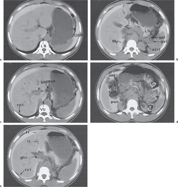
Fig. 25.1a–e CT anatomy of the peritoneal space and ligaments in the upper abdomen, as seen on five different levels. The patient has ascites; a water-soluble contrast medium has been administered, which helps to visualize the peritoneal space.


ass


falciform ligament


ghl


anterior subhepatic space


fl


gastrohepatic ligament


gpl


gastropancreatic ligament


gpplica


gastropancreatic plica


gsl


gastrosplenic ligament


GSR


gastrosplenic recess


Lr


lesser sac


lt


lateral recess of the lesser sac


LS


Morison pouch


pss


ligamentum teres


Mp


right coronary ligament


slrl


posterior subhepatic space


rcl


splenorenal ligament


Vb


vestibulum

Fig. 25.2 Topography of the posterior abdominal wall. Supramesocolic compartment. (Modified from Wegener.)


D


duodenum


GPP


gastropancreatic plica


GSL


gastrosplenic ligament


HDL


hepatoduodenal ligament with portal vein


LA


liver attachment


LS


lesser sac


LSS


left supramesocolic space


PCL


phrenicocolic ligament


RSS


right supramesocolic space


SHS


subhepatic space


TM


transverse mesocolon


Inframesocolic compartment.


AM


attachment of ascending mesocolon


DM


attachment of descending mesocolon


LIS


left infracolic space


LPG


left paracolic gutter


PD


pouch of Douglas


R


rectum


RIS


right infracolic space


RPG


right paracolic gutter


SM


root of the mesentery


U


uterus and adnexa


It extends from the proximal duodenum to the porta hepatis and contains the common hepatic duct, common bile duct, hepatic artery, and portal vein. It is a common route for spreading of carcinomas of the gallbladder and the biliary system.


The transverse mesocolon lies in the inframesocolic abdomen, contains middle colic vessels, and is a common site for spreading of pancreatic or colic processes.


The greater omentum is located anterior to the small bowel and represents the inferior continuation of the gastrocolic ligament. It is formed by a double reflection of the dorsal mesogastrium and has four layers of peritoneum.


The small bowel mesentery extends from the ligamentum of Treitz in the left upper quadrant down to the ileocecal valve, totaling a distance of ~15 cm. It contains superior mesenteric vessels.


The sigmoid mesocolon contains sigmoid and hemorrhoidal vessels and forms a pathway from the pelvis to the abdomen as it coalesces with the broad ligament in women. Infections and neoplasms of the sigmoid can spread not only to the pelvis, but also vice versa, with ovarian processes involving the sigmoid colon.


The broad and round ligaments serve as anterior suspenders of the uterus. The broad ligament contains uterine vessels branching from the internal iliac vessels, as well as lymph vessels and nerves. The ureters course distally through the base of the broad ligament. The lowermost portion of the broad ligament is called the cardinal ligament, which is the main supporter of the cervix and upper vagina. The round ligaments extend into the inguinal canal and farther down to the labia majora.


The medial umbilical ligament and the lateral umbilical folds are two ligament-like structures within the anterior wall of the abdomen. The latter contain hypogastric vessels.


Free intraperitoneal fluid can be seen on CT scans only in the presence of volumes > 50 mL. In the supramesocolic compartment, it initially collects within the Morison pouch and extends from there via the paravesical fossae into the paracolic gutters. In the inframesocolic compartment, fluid preferentially collects in the fossa of Douglas. In the presence of large amounts of as-cites, bowel loops typically are centrally positioned, and the mesentery can be clearly delineated.


Loculated (encapsulated) ascites indicates adhesions, whether benign or malignant. Thus, bowel loops do not float free within the central abdomen, but are displaced by loculated fluid. Unfortunately, CT attenuation of benign and malignant ascites is similar, thus rendering any CT-based differentiation impossible. Instead, distribution patterns may be suggestive. Unlike benign ascites, the amount of malignant ascites in the lesser sac typically matches the amount observed within the peritoneal cavity. Fluid predominantly confined to the lesser sac is more typically characteristic of diseases of the pancreas or abscesses of the lesser sac. Abscesses are loculated fluid collections with mass effect. CT attenuation usually is between 15 and 35 HU. The most sensitive CT feature is the presence of extraluminal gas, which is seen in only ~30% of all abscesses. (Loculated gas within any abdominal mass is always indicative of an abscess or necrosis formation.) A well-defined, hyperattenuating wall is a classic feature of a mature abscess.


CT is superior to plain film radiographs in detecting free intraperitoneal air, which usually collects ventrally in the midabdomen, beneath the abdominal musculature. Following surgical interventions, small amounts of free air may be detected up to 7 days on CT scans.


Relevant peritoneal and mesenteric processes can usually be identified on nonenhanced CT scans. Postcontrast scans are often useful to better delineate vessels and differentiate between solid and necrotic and cystic lesions. Irrespective of the scan technique, mesenteric abnormalities are detected much easier if the intestines are well opacified and thus in patients who previously received sufficient oral contrast. Usually patients should drink 1.5 L over a period of 60 to 90 minutes prior to the scan. On the CT table, rectal infusion of contrast media may further increase diagnostic accuracy. Table 25.1 lists the most relevant differential diagnoses of the peritoneum and mesentery.
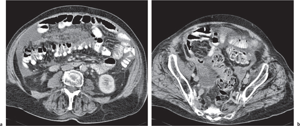
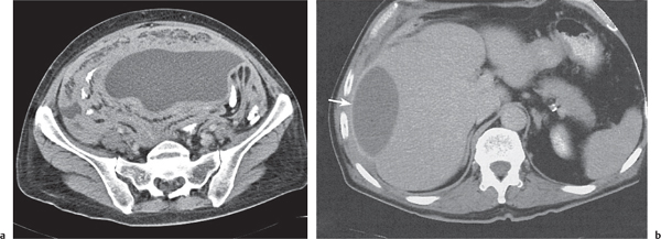
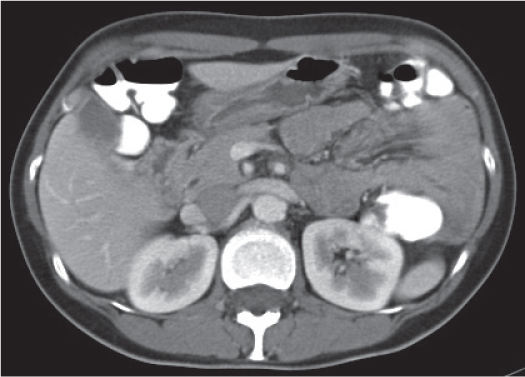
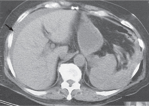
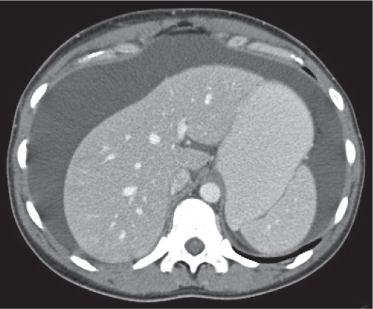
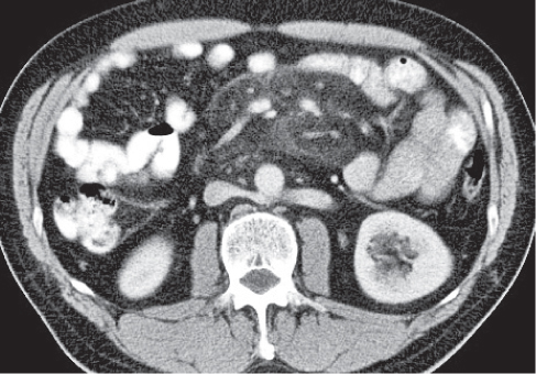
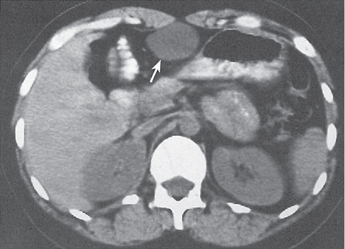
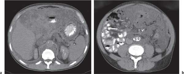
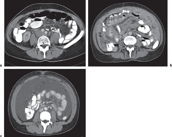
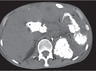


Table 25.1 Peritoneal, mesenteric, and omental collections and masses

Disease


CT Findings


Comments


Vascular


Omental infarction


Wedge-shaped to ovoid omental mass with hyper-attenuated streaks.


Scan recommendation:


Biphasic CT (nonenhanced CT, delayed phase [DP]).


Nonenhanced CT: Ill-defined, heterogeneous stranding of omental fatty tissue. Sometimes hyperdense free fluid collection.


DP: No enhancement.


Diagnostic pearls: Ill-defined mass with hyperdense stranding of omental fatty tissue in the right lower quadrant.


Typically located between the anterior abdominal wall and colon.


Congestion of omental fatty tissue due to a segmental infarction/ischemia.


Additional findings may be a focal inflammation with or without thickening of colonic and abdominal wall. Treatment: conservative or surgical excision (laparoscopy), depending on clinical presentation. Differential diagnosis: Acute appendicitis, epiploic appendagitis.


Infection


Peritonitis


Fig. 25.3a, b


Infectious, sometimes inflammatory affection of the peritoneum with or without the peritoneal cavity.


Scan recommendation:


Biphasic CT (nonenhanced CT, DP).


Nonenhanced CT: Hypodense intraperitoneal fluid collection. Hyperdense stranding and thickening of peritoneal fat.


DP: Slight hyperattenuation of soft tissue stranding and peritoneum.


Diagnostic pearls: Slight hyperattenuation of thickened peritoneum on postcontrast scans.


Arises from a posttraumatic or iatrogenic infection of the abdominal cavity.


Acute peritonitis: Intraperitoneal fluid, dilated mesenteric vessels, stranding of mesenteric fat, and enhancement of peritoneal membranes.


Chronic peritonitis: Extensive thickening of the peritoneum and adhesions (e.g., tuberculous peritonitis). Symptoms: Abdominal pain, fever. May lead to sepsis; thus, fast treatment is required: antibiotics, surgery.


Intraperitoneal abscess


Fig. 25.4a, b


Intraperitoneal fluid collections with mass effect.


Scan recommendation:


Biphasic CT (nonenhanced CT, DP).


Nonenhanced CT: Low-attenuation (10–40 HU) fluid collection often with gas bubbles.


DP: Strong rim enhancement.


Diagnostic pearls: Low-attenuation intraperitoneal fluid collection with rim enhancement and gas bubbles.


The surrounding peritoneal membrane becomes thicker and enhancing when the abscess matures.


Infected local peritonitis typically after perforation (ulcer, diverticulum), surgery, or inflammation (pancreatitis, pericholecystitis). Commonly found in the right subphrenic space, the subhepatic space, and the pouch of Douglas. Oral contrast, important to differentiate from fluid-containing bowel loops. Gas bubbles are pathognomonic but occur in 30% of cases.


Air–fluid levels may indicate a fistula to bowel loops. Treatment: Percutaneous abscess drainage (PAD) and antibiotics. Surgery is secondary treatment.


Tuberculosis (TB)


Fig. 25.4a, b


Mesenteric and omental masses and adenopathy.


Scan recommendation:


Biphasic CT (nonenhanced CT, DP).


Nonenhanced CT: Relatively high-density (15–30 HU), well-defined ascites.


DP: Hyperattenuation of thickened peritoneum.


Diagnostic pearls: Enlarged lymph may show rim enhancement.


Hematogenous disseminated TB from a distant focus (primary complex), usually from the lung. Rare, but often seen in immunocompromised patients.


Mesenteric lymphadenitis


Localized moderate enlargement of mesenteric lymph nodes associated with thickening of the wall of the distal ileum and cecum.


Scan recommendation:


Biphasic CT (nonenhanced CT, DP).


Nonenhanced CT: No additional information.


DP: Enhancement of lymph nodes and thickened bowel wall.


Usually caused by Yersinia enterocolitica, Campylobacter jejuni, or nontyphoid Salmonella species. Clinically, an important differential diagnosis of appendicitis and Crohn disease.


Internal hernias


Paraduodenal hernia (PDH)


Fig. 25.5


Cluster of dilated bowel loops with distorted mesenteric vessels.


Scan recommendation:


Biphasic CT (nonenhanced CT, DP).


Nonenhanced CT: Usually suffices for diagnosis. Oral contrast more important.


DP: Better delineation of distorted mesenteric vessels.


Diagnostic pearls: Cluster of dilated small bowel loops lateral to the ascending or descending duodenum.


Rare herniation of bowel loops through an acquired or congenital defect of the mesentery.


Right PDH (25%): Herniation through abnormal mesentericoparietal fossa of Waldeyer (lateral to ascending part of the duodenum).


Left PDH (75%): Herniation through abnormal paraduodenal mesenteric fossa of Landzert (next to the ligament of Treitz).


Transmesenteric hernia


Fig. 24.26 , p. 755


Cluster of dilated bowel loops with distorted mesenteric vessels.


Scan recommendation:


Biphasic CT (nonenhanced CT, DP).


Nonenhanced CT: Usually suffices for diagnosis. Oral contrast more important.


DP: Better delineation of distorted mesenteric vessels.


Diagnostic pearls: Dilated small bowel volvulus with twisted mesenteric vessels (“swirl” sign). Displacement of main mesenteric trunk to the left or right. Displacement of hepatic flexure inferiorly and posteriorly (common) or medial displacement of ascending/descending colon.


Typical postoperative complication after bowel surgery (Roux-Y, Whipple, etc.). In most cases, herniation of small bowel loops through artificial opening in mesentery of colon or small bowel (> 75% of cases through transverse mesocolon).


Trauma


Hematoma


Fig. 25.6


Local intraperitoneal or mesenteric fluid collection that otherwise mimics ascites.


Scan recommendation:


Multiphasic CT (nonenhanced CT, DP).


Nonenhanced CT: High-attenuating (30–80 HU).


DP: No enhancement.


Diagnostic pearls: Nonenhanced CT is important to avoid false-positive interpretation of high-attenuating blood as contrast media uptake. Attenuation may appear inhomogeneous due to incomplete coagulation.


Usually resulting from blunt trauma (particularly of the spleen and liver), bowel perforation with vascular erosion, spontaneous rupture of vascularized tumors (hepatoma, cavernous hemangioma, hepatic adenoma, angiosarcoma), extrauterine pregnancy, bleeding disorders, or excessive anticoagulation.


In chronic hematoma (> 1–2 weeks) attenuation drops to 0 to 20 HU. Treatment only in symptomatic patients (e.g., secondary infection, abscess formation, or bowel obstruction). PAD is treatment of choice.


Inflammation


Ascites


Fig. 25.7


Intraperitoneal fluid collection.


Scan recommendation:


Biphasic CT (nonenhanced CT, DP).


Nonenhanced CT: Low-attenuation fluid collection (< 30 HU). Density increases with protein content. Typical ascites: bile < 20 HU; urine ~10 to 15 HU; chylous fluid < 0 HU.


DP: Rim enhancement indicates concomitant perito-neal inflammation and abscess formation.


Diagnostic pearls: Loculation (nonuniform distribution) of ascites indicates postoperative, inflamma-tory, or neoplastic adhesions. Dense (> 30 HU) ascites suggests intraperitoneal hemorrhage or TB. The lesser sac usually is only moderately filled. Large amount of ascites in the lesser sac, small nodules on the smooth surfaces of the liver and spleen, and mesenteric lymph nodes are characteristic of peritoneal carcinomatosis.


Ascites may develop from right heart failure, abdominal vein occlusion, constrictive pericarditis, hypoalbuminemia, liver failure, myxedema, peritonitis, pancreatitis, intestinal perforation, glomerulonephritis, peritoneal carcinomatosis, and malignant lymphomas (blockage of lymph flow).


The combination of fibrous ovarian tumors, hydrothorax, and benign ascites is called Meigs syndrome. Discrete ascites appears either as a hypodense rim around intraperitoneal organs (particularly the right liver lobe and the spleen) or small fluid collections in the Morison pouch, perihepatic, or Douglas space. Larger amounts of ascites lead to fluid collection in paracolic gutters and centralization of small bowel loops in the uppermost part of the peritoneal cavity.


Biliary ascites, biloma


Well-defined, low-density fluid collection in the left or right supramesocolic space.


Scan recommendation:


Multiphasic CT (nonenhanced CT, hepatic arterial phase [HAP], portal venous phase [PVP], DP). Nonenhanced CT: Low-density fluid collection (~20 HU) DP: Rim enhancement indicative of infection/inflammation.


Usually iatrogenic or posttraumatic, rarely spontaneous rupture of the gallbladder. Bile causes local inflammation, sealing the fluid collection near the porta hepatis (i.e., biloma).


Pancreatic ascites


Fig. 22.10a–c , p. 725


Diffuse, low-density fluid collection in peripancreatic fatty tissue, lesser sac, and anterior pararenal space (i.e., ventral to fascia of Gerota).


Scan recommendation:


Multiphasic CT (nonenhanced CT, HAP, PVP, DP). Nonenhanced CT: Sufficient for visualization of ascites. DP: Additional information of pancreatic perfusion (necrosis).


Typically observed in patients with pancreatitis, after seat-belt injury (disruption of pancreas corpus [spine as hypomochlion, or center of rotation]), or following pancreas surgery.


Fibrosing mesenteritis (retrac-tile mesenteritis, mesenteric panniculitis, mesenteric lipodystrophy)


Fig. 25.8


Separation of bowl loops with kinking and angulation, mesenteric thickening with fine stellate pattern.


Scan recommendation:


Biphasic CT (nonenhanced CT, DP).


Nonenhanced CT: May contain calcified or enlarged mesenteric lymph nodes and central necrotic cysts. DP: Visualization of encased mesenteric vessels. May show slightly enhancing pseudocapsule.


Diagnostic pearls: Mesenteric stellate mass encasing mesenteric vessels with a preserved hypodense (fat) perivascular halo.


Inflammation, fibrosis, and fatty infiltration of the mesentery.


Usually located in the root of small bowel mesentery, less often in mesentery root of the colon, the omen-tum, or within the retroperitoneum (peripancreatic tissue). Associated with Gardner syndrome and fibrosing mediastinitis.


Multiple mesenteric soft tissue masses represent fibromas.


Panniculitis is regarded as an early stage of fibrosing mesenteritis. Typically appears as ill-defined soft tissue stranding of mesenteric fat.


Benign neoplasms


Pancreatic pseudocyst


Well-defined, near-water-density (retro-) peritoneal or mesenteric mass.


Scan recommendation:


Biphasic CT (nonenhanced CT, DP).


Nonenhanced CT: Circumscript near-water-density (< 15 HU) mass.


DP: Often distinct rim enhancement.


See also Fig. 22.11 , p. 725 .


The most common lesion of the lesser sac. May invade the mesentery.


Likely only in patients with an acute or chronic pancreatitis.


Treatment of choice in symptomatic patients is transgastral/percutaneous drainage.


Mesenteric cyst (omental cyst, duplication cyst, lymphangioma)


Fig. 25.9


Usually near-water-density, single or multilocular cysts up to several centimeters in size.


Scan recommendation:


Multiphasic CT (nonenhanced CT, DP).


Nonenhanced CT: Attenuation between chylous (< 0 HU) and blood (> 40 HU), depending on main cyst content. In chronic cases, rim calcifications.


DP: Sometimes discrete rim enhancement.


Diagnostic pearls: Low-attenuation, fluid-filled intra-abdominal cyst with thin rim and subtle internal septa.


Hamartomatous lesions lined by mesothelial cells with chylous, serous, or occasionally hemorrhagic fluid contents. Usually asymptomatic.


Omental cysts may be pedunculated.


In some cases, a water–fat level is present. Differential diagnoses include pancreatic (pseudo-) cyst, cystic mesothelioma, and cystic teratoma.


Fibromatosis (desmoid tumor)


Well-defined, usually large encapsulated homogeneous mass of variable attenuation and enhancement.


Scan recommendation:


Biphasic CT (nonenhanced CT, PVP).


Nonenhanced CT: Isodense to muscle.


PVP: Usually discrete enhancement. Optimal for visualization of encased mesenteric vessels.


Histologically, an aggressive tumor composed of fibroblastic tissue.


Fibromatosis is the most common primary tumor of the mesentery. Known association with local trauma, Gardner syndrome, estrogen therapy, and surgery. Typically involves small bowel mesentery and leads to displacement and obstruction of bowel loops. Infiltration into adjacent organs/muscles is observed.


Lipoma


Well-defined, marginated, nonenhancing, fat-density mass.


Scan recommendation:


Multiphasic CT (nonenhanced CT, DP).


Nonenhanced CT: Low attenuation (~10–15 HU with minimal values below 0 HU).


DP: No enhancement.


Diagnostic pearls: Low-attenuation framed lesion without internal septations.


The second most common primary benign solid tumor of the mesentery.


Neurofibroma


Well-defined, low-density mesenteric mass.


Scan recommendation:


Biphasic CT (nonenhanced CT, DP).


Nonenhanced CT: Well-defined hypoattenuating mass.


DP: Homogeneous enhancement.


Histologically, lesions are built of endoneural myxoid matrix.


May arise as isolated lesions or be part of a clinically proven neurofibromatosis.


Splenosis


Fig. 21.7a, b , p. 713


Single or multilocular intra-abdominal soft tissue masses.


Scan recommendation:


Biphasic CT (nonenhanced CT, DP).


Nonenhanced CT: Well-defined spleenlike attenuation.


DP: Homogeneous spleenlike attenuation.


Diagnostic pearls: Single or multilocular nodules on the serosal surface of the small bowel, greater omentum, or parietal peritoneum with spleenlike attenuation.


Autotransplantation of splenic tissue after splenic trauma or splenectomy.


Usually asymptomatic, but may cause abdominal pain or gastrointestinal hemorrhage.


Castleman disease (angiomatous lymphoid hamartoma)


Large homogeneous, well-defined mass.


Scan recommendation:


Multiphasic CT (nonenhanced CT, HAP, PVP, DP). Nonenhanced CT: Well-defined, isodense to muscle. DP: Moderate contrast enhancement.


Two histologic subtypes: Hyaline-vascular (90%) and plasma cell type (10%). Benign massive enlargement of lymph nodes. Most (70%) occur in the mediastinum of young healthy people, but also in the mesentery and omentum.


Malignant neoplasms


Pseudomyxoma peritonei


Fig. 25.10a,b


Diffuse, multiseptated fluid collection with loculations.


Scan recommendation:


Multiphasic CT (nonenhanced CT, DP).


Nonenhanced CT: Multiple grapelike, hypodense (15–30 HU) intra-abdominal cysts. Cyst walls partly calcified.


DP: Hyperattenuating, thickened peritoneal and omental surface.


Diagnostic pearls: Multiple grapelike hypodense cysts with rim enhancement, scalloping of liver/spleen surface, and displacement of intestinal structures and mesentery. See also Fig. 29.62 , p.832.


Rare disease. Histologically, an intraperitoneal accumulation of gelatinous, mucinous material, which usually derives from the spread of ovarian mucinous cystadenocarcinoma. Malignant degeneration of an appendicular mucocele is a less common cause. Other (rare) origins include stomach, colon, uterus, bile ducts, and pancreas.


Contrast media enhances solid components and improves the visibility of the septations. Mucinous components remain hypoattenuating.


Lymphoma


Fig. 24.22a, b , p. 753


Well-defined soft tissue density masses > 10 mm in size.


Scan recommendation:


Multiphasic CT (nonenhanced CT, DP).


Nonenhanced CT: Isodense to muscle.


DP: Homogeneous enhancement.


Diagnostic pearls: Well-defined, partly coalescing homogeneous enhancing masses.


The most common neoplastic mesenteric mass. Predominantly non–Hodgkin lymphomas (61%), Hodgkin disease involves the mesentery in only 5% of cases.


Lymphomas typically encase instead of invade mesenteric vessels, which may result in a “sandwich sign.”


Peritoneal carcinomatosis (PC)


Fig. 25.11a–c


Any combination of ascites, nodular omental thickening, mesenteric stranding, scalloping of parenchymal organ’s contours, and enlarged mesenteric lymph nodes.


Scan recommendation:


Biphasic CT (nonenhanced CT, DP).


Nonenhanced CT: Lymph nodes, ascites, and omentum hypoattenuating.


DP: Enhancement of peritoneal/omental nodularities.


Diagnostic pearls: Omental caking, bowel obstruction (subileus), and enlarged mesenteric lymph nodes.


Appearance of PC ranges from a thickened nodular peritoneal surface to a cakelike omental mass. Peritoneal metastases most commonly derive from ovarian, colonic, or gastric cancer. Calcification and cystic lesions on liver/spleen contour or peritoneal surface suggests ovarian cystadenocarcinoma. Ascites presents in > 70% and may be loculated. Omental fat is involved in > 70% and appears normal, permeated with multiple discrete enhancing nodules, or as diffuse caking (omental caking). Mesenteric involvement is present in > 60% mainly as diffuse or stellate soft tissue stranding. Diagnostic accuracy of CT in the diagnosis of PC is low.


Malignant mesothelioma (MM)


Peritoneal associated with calcified pleural plaques.


Scan recommendation:


Biphasic CT (nonenhanced CT, DP).


Nonenhanced CT: Subtle to distinct calcified plaques. DP: Homogeneous enhancement.


Diagnostic pearls: Stellate peritoneal mass or omental caking, infiltrating vessels and adjacent viscera. Calcifications are present in the majority of cases. Disproportionately little ascites in 90%.


MM is associated with excessive exposure to asbestos (~20–40-y latent period). Twenty-five percent of MM cases arise from the peritoneum. Three histologic subtypes:


1. Carcinomatous type (most common): Diffuse thickening of the peritoneum, multiple mesenteric and peritoneal nodules, and fixation of the small bowel


2. Sarcomatous type (20%): Large encapsulated mass with local invasion


3. Mixed type


Liposarcoma


Fig. 25.12


Nodular myxoid mass with regions of hemorrhage and necrosis.


Scan recommendation:


Biphasic CT (nonenhanced CT, DP).


Nonenhanced CT/DP: Appearance variable, depending on main tissue component. Often multiple heavily calcified lesions.


Most common in the retroperitoneum, rare in the omentum or mesentery.


Five histologic subtypes: pleomorphic, round cell, myxoid, sclerosing, and lipomalike. Often indistinguishable from lipoma.


Spindle cell tumors (leiomyoma, leiomyosarcoma)


Large heterogeneous, solid mass typically with cystic spaces representing necrosis.


Scan recommendation:


Multiphasic CT (nonenhanced CT, HAP, PVP, DP).


Nonenhanced CT: Appearance variable, depending on main tissue component.


DP: Intense contrast enhancement of the solid components.


The most common primary omental tumor. Leiomyosarcoma is more common than leiomyoma.

Fig. 25.3a, b Peritonitis. Soft tissue stranding of mesentery fat (a) and slight hyperattenuation of thickened peritoneum (b) on postcontrast CT scans.
Fig. 25.4a, b Intraperitoneal abscesses. Mesenteric abscess presenting as a low-fluid collection with rim-enhancement in a patient with known tuberculosis (a). Subphrenic abscess (b). A lesion with similar CT appearance is located between the liver and right hemidiaphragm (arrow).
Fig. 25.5 Paraduodenal hernia to the left. Cluster of poorly opacified small bowel loops lateral to the well-opacified descending duodenum.
Fig. 25.6 Intraperitoneal hematoma due to hepatic laceration. A hyperdense crescent perihepatic lesion is visible on noncontrast scan.
Fig. 25.7 Ascites. Near-water density surrounding the liver, stomach, and spleen.
Fig. 25.8 Panniculitis. Discrete localized soft tissue stranding within the mesenteric root simulating a slightly enhancing pseudocapsule and encasement of mesenteric vessels.
Fig. 25.9 Mesenteric cyst. Smooth, thin-walled, near-water-density collection between the greater curvature and abdominal wall (arrow).
Fig. 25.10a, b Pseudomyxoma peritonei. Diffuse, multiseptated intra-abdominal fluid collection with loculations.
Fig. 25.11a–c CT appearance of peritoneal carcinomatosis. Findings range from subtle mesenteric soft tissue stranding (a) to a cakelike omental mass (b). In ovarian cancer (see also Fig. 19.25d), the only finding may be marked ascites without nodular mesenteric thickening (c).
Fig. 25.12 Peritoneal liposarcoma presenting with multiple heavily calcified lesions.

Only gold members can continue reading. Log In or Register to continue

Stay updated, free articles. Join our Telegram channel

Jul 6, 2020 | Posted by in GENERAL RADIOLOGY | Comments Off on 25 Peritoneum and Mesentery

Full access? Get Clinical Tree
Get Clinical Tree app for offline access