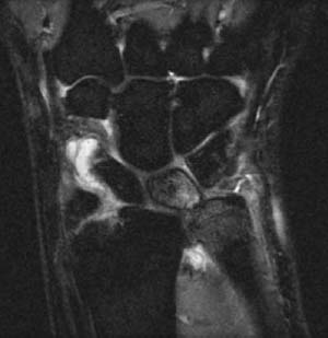
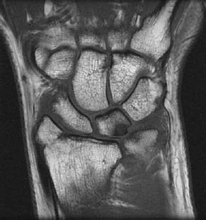
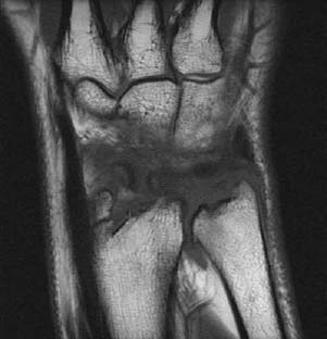
CASE 25 Peter L. Munk and Anthony G. Ryan A 46-year-old man presented with chronic pain on the inside of his wrist, exacerbated while performing home improvements, particularly when hammering and when lifting his toolbox by the handle. Years previously he had sustained a fracture of the distal radius. Figure 25A Figure 25B Figure 25C A coronal STIR image (Fig. 25A) shows high signal intensity in the head of the ulna and in the ulnar aspect of the lunate. The distal lateral aspect of the ulnar head abuts the lunate. There is loss of contiguity of the radial and ulnar carpal articular surfaces. An essentially free portion of the triangular fibrocartilage (TFC) is seen just distal to the ulnar head. A bony fragment is projected distal to the distal radius in the expected position of the radial styloid. A ganglion is seen in relation to the radial aspect of the scaphoid. A coronal T1-weighted image (Fig. 25B) volar to the STIR image shows a low signal intensity focus on the ulnar aspect of the lunate and a corresponding low signal intensity focus on the adjacent ulnar head. Bony irregularity of the radial styloid process is evident. A coronal T1-weighted image (Fig. 25C) dorsal to the STIR image shows an irregular radial styloid process, distal radius, and ulna, as well as an osteophyte on the ulnar aspect of the distal radioulnar joint. Ulnar impaction syndrome with TFC complex tear. Proper functioning of the wrist requires smooth articulation of the proximal carpal row with the distal radius and ulnar. The TFC complex plays a crucial role in this regard. Most commonly, patients demonstrate what is referred to as positive ulnar variance (although this is not a sine qua non for degenerative ulnar impaction syndrome). Ulnar variance refers to the relative lengths of the distal articular surfaces of the radius and ulna. With positive ulnar variance, the ulna is longer than the radius; in particular, the carpal articular surface of the ulnar projects more distally than the radial carpal articular surface. Because ulnar variance can depend on the position of the wrist, that is, supination versus pronation, variance is usually measured in neutral rotation using a posteroinferior (PA) projection, the elbow flexed to 90 degrees and the shoulder abducted by 90 degrees. When present, pronation of the forearm and gripping both exacerbate the degree of positive variance.
Triangular Fibrocartilage Tear
Clinical Presentation


Radiologic Findings
Diagnosis
Differential Diagnosis
Discussion
Background
Etiology
Stay updated, free articles. Join our Telegram channel

Full access? Get Clinical Tree


