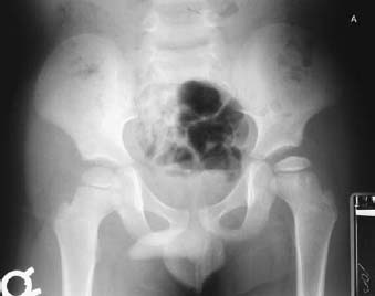
PART II Congenital and Pediatric Conditions Anthony G. Ryan, Peter L. Munk, and Laurel O. Marchinkow A 5-year-old boy presented with a 3-month history of a limp. Figure 26A A flattened irregular left capital femoral epiphysis is shown in Fig. 26A. Legg-Calvé-Perthes disease. Otherwise known as coxa plana, Legg-Calvé-Perthes disease is avascular necrosis of the femoral head in children, most common between the ages of 4 and 8, with a male (5:1) and Caucasian preponderance. The condition occurs bilaterally in 15%. The condition is most commonly idiopathic, but in 30% of cases, it is secondary to trauma. Hip pain and limp, lasting between 1 week and 6 months (mean = 2.7 months). Early severe degenerative hip joint disease. Legg-Calvé-Perthes disease is one of a heterogeneous group of disorders known as the osteochondritides, characterized radiologically by fragmentation, collapse, sclerosis, and eventual reossification of the involved bone, and pathologically by osteonecrosis. The femoral head is at particular risk for this latter process by virtue of the tenuous blood supply to the femoral epiphysis via the medial circumflex and lateral epiphyseal arteries. If the blood supply is disrupted by trauma or other causes (e.g., intravascular), 6 to 12 hours of anoxia leads to death of the hematopoetic cells. Twelve to 48 hours of anoxia is required for bone cell death. Forty-eight to 60 hours of anoxia leads to death of the marrow fat cells. This results in loss of fat cell nuclei and the formation of fat cysts, both of which are taken as pathological features of established osteonecrosis. Surrounding the central zone of cell death is a zone of ischemic injury, and at the margin of this is an advancing hyperemic zone as a result of recanalization of thrombosed vessels and neovascularity. The radiologic findings parallel the evolution of the disease process. Thus, at the time of cell death, conventional radiographs will be normal, as will radionuclide studies.
CASE 26
Legg-Calvé-Perthes Disease
Clinical Presentation
Radiologic Findings
Diagnosis
Discussion
Background
Etiology
Clinical Findings
Stages of Disease
Complications
Pathology
Imaging Findings
RADIOGRAPHY
Related posts:
Stay updated, free articles. Join our Telegram channel

Full access? Get Clinical Tree


