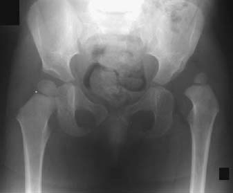
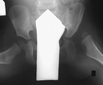
CASE 27 Anthony G. Ryan and Peter L. Munk A 21-month-old girl was noted to have a waddling gait and, on physical examination, was found to have a shorter left leg. Figure 27A Figure 27B The anteroposterior (AP) (Fig. 27A) and Von Rosen (with 45-degree abduction and internal rotation) (Fig. 27B) projections show Putti’s triad: the left proximal femur is dislocated superolaterally, the acetabular angle is steep, and the capital femoral epiphysis is smaller than on the contralateral side. Congenital dysplasia of the hip. This is a relatively common condition, affecting 0.15% of newborns, with a marked female (8:1) and left-sided (11:1) preponderance. In the majority of cases, the cause is a combination of ligamentous laxity (secondary to maternal estrogens) and acetabular dysplasia, whereby the acetabulum is steeper and more shallow than normal. The femoral head is thus predisposed to fall out of the acetabulum, putting the hip at risk of subluxation/dislocation when exposed to mechanical stress, for example, breech birth. The hip is “dislocatable” on provocation, or the hip is already dislocated. When the hip is dislocatable, the head of the femur jerks in and out of the acetabulum with posterior pressure applied along the length of the femur. When already dislocated, a positive Ortolani’s click describes the snapping of the femoral head back into the acetabulum. If relocation is not possible, the Ortolani will be falsely negative, in which case other signs may be indicative of the condition: If the child is walking, the gait will be abnormal, with a positive Trendelenburg’s sign: the contralateral pelvis drops when the child stands on the affected limb.
Congenital Dislocated Hip
Clinical Presentation
Radiologic Findings
Diagnosis
Discussion
Clinical Findings
Imaging Findings
RADIOGRAPHY
Related posts:
Stay updated, free articles. Join our Telegram channel

Full access? Get Clinical Tree


