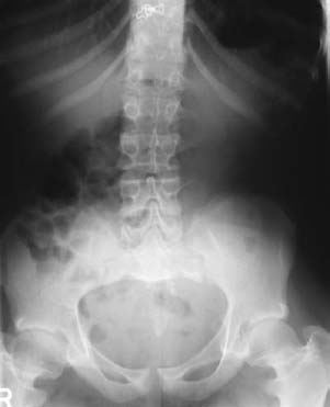
CASE 28 Brian Edward Reeves, Anthony G. Ryan, Peter L. Munk, and Thomas Pope A 16-year-old girl presented with back pain. Physical exam revealed that she has short arms and legs and macrocephaly, and she is bowlegged. Figure 28A A lumbar spine anteroposterior (AP) radiograph (Fig. 28A) shows lumbar spinal stenosis with a gradual decrease in interpedicular width cranially to caudally. Squaring of the iliac wings and bilateral coxa vara deformities are also seen. The femoral necks are short and thick bilaterally. Achondroplasia. Although other rhizomelic short-limbed dwarf syndromes may have similar radiographic findings, the combination of the typical phenotype and radiographic findings makes no other diagnosis admissible. Achondroplasia, affecting ~1 in every 25,000 to 40,000 births, is the most common short-limbed dwarf syndrome. The characteristic features of achondroplasia are present at birth, and diagnosis is made by physical examination and the characteristic skeletal radiographic findings. Affecting all races and both sexes, achondroplasia is transmitted as an autosomal dominant disorder with complete penetrance. Eighty percent of achondroplasia cases are caused by mutations in the gene for fibroblast growth factor receptor-3, whose primary function is to limit osteogenesis. These mutations severely limit enchondral ossification. Advanced paternal age (> 40 years old) is identified as a major risk factor. Achondroplasia is due to a defect in enchondral bone formation, with a resulting decrease in the rate at which epiphyseal cartilage cells convert to bone. This results in shortening of the long bones and a decrease in vertebral body and posterior arch diameters. Gross motor development is frequently delayed, and head control, standing, and ambulation can lag behind by 6 months. Speech and language problems may occur because of the abnormal maxillomandibular relationship. Cognitive skills are preserved. Physical examination findings may include macrocephaly, frontal bossing, flattening of the nasal bridge, midface hypoplasia, and prominent mandibles. Recurrent otitis media is common due to poor drainage of the eustachian tube from underdevelopment of the midface, hypertrophy of the tonsils, and temporal bone abnormalities.
Achondroplasia
Clinical Presentation
Radiologic Findings
Diagnosis
Differential Diagnosis
Discussion
Background
Etiology
Pathophysiology
Clinical Findings
Related posts:
Stay updated, free articles. Join our Telegram channel

Full access? Get Clinical Tree


