CASE 29 Anthony G. Ryan and Peter L. Munk A child presented with increasing irritability over the previous several weeks with marked swelling and tenderness in both lower extremities. Birth history was unavailable, as the child was adopted shortly after birth. Clinical examination demonstrated pain in both lower extremities affecting both thighs and legs. Examination was otherwise unremarkable apart from bilateral sport affecting the sclera of both eyes. Figure 29A A frog-leg view of the hips and thighs (Fig. 29A) shows bilateral partially healed fractures. Two fractures are noted on the left with a single fracture distally on the right. The fractures appear subacute. Osteogenesis imperfecta. Osteogenesis imperfecta describes a group of genetic conditions of varying severity resulting in multiple pathologic fractures in the fetus, neonatal period, or childhood, the diagnosis of which is made from clinical, genetic, and radiographic features. Radiologists are frequently called as expert witnesses and asked to distinguish osteogenesis imperfecta from child abuse to avoid an improper accusation of child abuse in a child with obvious osteogenesis imperfecta. This disorder of connective tissue, found in 1 in 40,000 births, affects the synthesis and quality of fibrillar collagen, resulting in congenital osteoporosis secondary to inadequate osteoid formation with normal mineralization. Inadequate amounts of osteoid are produced to balance physiological osteolysis. The bones are osteoporotic, gracile, and, consequently, extremely fragile, causing bowing and fractures. Excessive callus formation is characteristic and may involve the entire length of the bone. Some fractures may heal with a pseudoarthrosis. Depending on the subtype, at presentation deformities from previous fractures may be dominant, or the bones may be bowed secondary to microfractures. Osteogenesis imperfecta may be diagnosed antenatally by chorionic villus sampling. Also known as osteogenesis imperfecta tarda, patients are typically of normal birth weight and length. The first fracture typically occurs in the second or third year of life, although presentation may be as early as delivery (intrapartum fractures in 20%) or as late as 6 years of age. On examination, blue sclerae (found in 90%) and presenile deafness (20% secondary to otosclerosis and auditory nerve impingement) will be seen. There are variable dental findings (30%) secondary to dentin dysplasia. Joint hypermobility may be evident secondary to ligamentous laxity. These children are frequently stillborn or die soon postnatally secondary to pulmonary hypoplasia. This is the mildest form with the best prognosis. The condition is classified into four major types.
Osteogenesis Imperfecta
Clinical Presentation
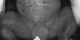
Radiologic Findings
Diagnosis
Differential Diagnosis
Discussion
Background
Etiology
Pathophysiology
Clinical Findings
Type 1
Type 2
Type 3
Type 4
Stages of Disease
Related posts:
Stay updated, free articles. Join our Telegram channel

Full access? Get Clinical Tree


