29 Pelvis


10.1055/b-0034-75822

29 Pelvis

Dieudonné\, Grégory and Burgener\, Francis A.

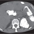
An understanding of soft tissue and bony pelvic anatomy on computed tomography (CT) aids in the detection of pelvic diseases and recognition of their patterns of spread. Nearly all major organ systems are represented in the pelvis, including the gastrointestinal (GI), genitourinary, central and peripheral nervous, angiolymphatic, and musculoskeletal systems. The limits of the pelvis are defined by the innominate bones, sacrum, and coccyx. The internal and external pelvic muscular and bony frameworks are relevant, as disease processes arising in these structures sometimes manifest within the pelvic cavity (Figs. 29.1 , 29.2).


The pelvic peritoneal reflections delineate significant but not insurmountable boundaries between extraperitoneal structures and the peritoneal cavity. The peritoneal reflections over the pelvic organs, vessels, and ligaments define several peritoneal pelvic cavity recesses, the largest and most gravity-dependent of which is the rectovesicle pouch. The rectovesicle pouch, along with the pelvic portion of the greater omentum, is one of the most common sites of tumor implants in peritoneal carcinomatosis.


Pelvic peritoneal reflections often form anatomical landmarks recognizable on CT images where they envelop organs and the associated vascular and lymphatic systems. The peritoneal reflection forming the sigmoid mesocolon encompasses a portion of the proximal rectum and sigmoid colon, along with their associated angiolymphatic structures. The sigmoid mesocolon is more easily seen in the presence of ascites (Fig. 29.3). In addition, the small bowel and its mesentery, as well as their associated pathologic processes, often extend into the pelvis (Fig. 29. 4 ).


In women, the broad ligament is that part of the peritoneal reflection surrounding the margins of the uterus, fallopian tubes, and proximal round ligaments extending to the pelvic sidewall. The ovary is attached to the posterior broad ligament by the mesovarium. The peripheral broad ligament from the ovary to the pelvic sidewall that contains the ovarian vessels is referred to as the suspensory ligament of the ovary. The uterus divides the rectovesical pouch into the anterior vesicouterine pouch and the posterior rectouterine pouch, also known as the cul-de-sac or pouch of Douglas.

Fig. 29.1 Abdominal wall desmoid. Heterogeneously enhancing soft tissue mass in the anterior abdominal wall with impression onto the colon.

Although the remaining pelvic structures, such as the rectum, anus, distal ureters, bladder, cervix, vagina, seminal vesicles, prostate, and sidewall structures, do not readily protrude into the pelvic peritoneal cavity, they are often associated with fascial planes that frequently confine pathologic processes. These boundaries localize lesions to their site of origin, guiding the differential diagnosis of a pelvic mass.


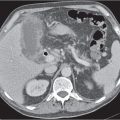
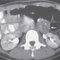
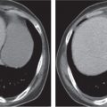
Ectopic organs, such as pelvic kidneys (Fig. 29.5), are readily identified on CT. Pelvic testicles present as elliptical foci along the course of the testicles’ descent, frequently within the inguinal canal (Fig. 29.6).


Some surgical procedures may result in pseudolesions due to the displacement of normal pelvic structures. In particular, abdominoperineal resection of the rectum can result in a posterior presacral position of the uterus in women or the prostate and seminal vesicles in men (Fig. 29.7). These normal presacral masses must be distinguished from presacral recurrent tumor and postoperative fibrosis. If a suspicious mass is encountered, biopsy is warranted. If there is question that a presacral mass is a displaced normal structure such as the seminal vesicles or uterus, magnetic resonance imaging (MRI) is often helpful.


Midline or lateral surgical transposition of the ovaries in women can also be problematic if the history of this procedure is not elicited, especially if there is a superimposed functional cyst. Lateral surgical ovarian transposition procedures are sometimes performed to preserve ovarian function in young women with cervical carcinoma who will receive radiation therapy. The ovaries are placed in the iliac fossae or high in the lateral abdomen (Fig. 29.8). Midline ovarian transposition may be performed in young women who will receive pelvic nodal radiation for lymphoma.


Other iatrogenic “pseudolesions” are intestinal urinary reservoirs in patients with urinary diversions, tissue expanders to displace bowel from radiation ports prior to radiation therapy (Fig. 29.9), and implanted reservoirs for penile prostheses and artificial sphincters.


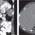
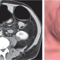
Renal and pancreatic transplants are usually placed within the pelvis (Fig. 29.10). Although they are typically imaged with ultrasound, CT is occasionally performed. The pancreatic transplant is often connected to the bladder by a segment of bowel to allow drainage of exocrine fluids. Care must be taken to avoid confusing this with an abnormal collection. Renal and pancreatic peritransplant collections, such as hematomas, seromas, abscesses, urinomas, lymphoceles, and pancreatic pseudocysts, are readily detected with CT.


The accompanying tables of differential diagnoses of pelvic pathologic processes are predominantly organized by organ of origin. However, because the exact site of origin is not always evident, tables relating to a variety of lesions disseminating within the peritoneal cavity and originating from the pelvic cavity walls are also included. Table 29.1 discusses pelvic GI lesions, Table 29.2 lesions of the female and male pelvis, Table 29.3 urinary bladder lesions, and Table 29.4 peritoneal and extraperitoneal lesions.

Fig. 29.2 Muscular dystrophy. Diffuse fatty replacement of muscles.
Fig. 29.3a, b Sigmoid mesocolon. The sigmoid colon and its mesentery (arrows), referred to as the sigmoid mesocolon, are suspended by pelvic ascites in this patient with carcinomatosis (a). Distended air–fluid-filled sigmoid tapering to a beak (arrow) without perceived wall thickening/mass. Note the collapsed twisted segment (arrowheads) of the distal sigmoid (sigmoid volvulus: twisting of the mesocolon about its mesenteric axis) (b).
Fig. 29.4 Mesenteric adenopathy. Multiple soft tissue attenuation nodes distributed within the small bowel mesentery (arrows); contrast this with the nonopacified bowel segments within the left anterior hemipelvis.
Fig. 29.5a, b Congenital pelvic kidney. Axial (a) and parasagittal (b) views showing the ectopic location of the left kidney within the pelvis.
Fig. 29.6 Undescended testis. The un-descended testicle (arrow) in this patient is within the left inguinal canal.
Fig. 29.7 Pelvis following abdominoperineal resection for rectal carcinoma. Normal presacral prostate position (star) following abdominoperineal resection. There is also partially calcified left obturator adenopathy (arrow) due to recurrent tumor.
Fig. 29.8 Ovarian transposition. The ovaries (arrows) were surgically placed in the lateral abdomen in this patient undergoing pelvic radiation for cervical carcinoma.
Fig. 29.9 Tissue expander. Surgically placed expandable device (arrows) that is used to displace the bowel from the area of a radiation port.
Fig. 29.10 Renal transplant. Kidney within the anterior right hemipelvis. Note the surgical clips, focus of calyceal gas (arrow). This patient has a filling defect (thrombus) within the left common iliac vein.


Table 29.1 Pelvic gastrointestinal lesions

Disease


CT Findings


Comments


Carcinoma


Fig. 29.11


Fig. 29.12


Segmental wall thickening or polypoid luminal masses. Search for lymph nodes, hepatic metastases, and peritoneal or omental tumor implants.


Often difficult to distinguish bowel contractions and luminal fecal material from focal lesion. Rescan area of concern or barium enema versus small bowel follow-through may be necessary to confirm in the absence of associated secondary signs of tumor (e.g., perienteric fat infiltration).


Lymphoma


Fig. 29.13


Segmental or diffuse, irregular or smooth bowel wall thickening, sometimes associated with luminal expansion. May occur with or without adenopathy.


These are typically non–Hodgkin lymphomas. Gastrointestinal (GI) lymphoma may also be encountered in patients positive for human immunodeficiency virus (HIV) and in posttransplant lymphoproliferative disorder.


Mesenchymal neoplasms


Fig. 29.14


Polypoid luminal or exophytic intermediate-attenuation mass causing luminal narrowing. The presence of calcifications would favor this diagnosis. Lipomas (see also Fig. 29.25) typically are polypoid and usually have uniform fat attenuation.


Other types of mesenchymal neoplasms are leiomyomas, leiomyosarcomas, and carcinoid tumors. Cystic necrosis is often seen in leiomyosarcomas. Carcinoid tumors may have associated mesenteric fibrotic response, which can lead to intestinal ischemia.


Metastases


Fig. 29.15a, b


Soft tissue nodules, adjacent to or impinging on the small bowel lumen. Scirrhous lesions, intramural deposits, or polypoid intraluminal masses may be present. The greatest portion of the metastatic deposit is located in the mesentery adjacent to the wall of the small bowel. An omental “cake” may be recognized as an extensive soft tissue mass separating the colon or small bowel from the anterior abdominal wall.


Most common primary tumors are carcinomas of lung, breast, colon, pancreas, kidney, uterus, and skin.


Diverticulitis


Fig. 29.16


Segmental wall thickening with or without pericolonic infiltrative changes, fistulas, and/or abscesses. Usually occurs in patients with visible diverticula.


Majority of cases involve sigmoid colon, rarely other colonic sites or small bowel diverticula.


Inflammatory bowel disease


Fig. 29.17


Fig. 29.18


Fig. 29.19


Segmental or diffuse, irregular or smooth bowel wall thickening, with or without perienteric infiltration.


Examples include Crohn disease, which may involve several bowel segments, with or without fistulas; typically spares rectum. May also observe associated mesenteric fibrofatty proliferation. Graft versus host disease can lead to fold and wall thickening and can be associated with pneumatosis intestinalis. Radiation enteritis can be acute and present within a few weeks of therapy, or chronic, manifesting a few months or more after therapy. Acute effects are due to direct cellular toxicity and chronic effects from a radiation-induced vasculitis.


Infectious enterocolitis


Fig. 29.20


Segmental or diffuse, irregular or smooth bowel wall thickening. Nonneoplastic bowel wall thickening will often result in a low- (near-water) attenuation value at the sites of involvement.


Examples include pseudomembranous colitis, which may produce marked wall thickening, and other microbial agents, such as cytomegalovirus, which is a common HIV-related infection.


Ischemia


Fig. 29.21


Fig. 29.22


Fig. 29.23a–c


Segmental or diffuse, irregular or smooth bowel wall thickening with or without pneumatosis intestinalis.


Survey mesenteric arteries and veins for the presence of thrombi and/or gas. Ischemia typically spares the rectum because of its dual blood supply.


Endometriosis


Mixed or uniform intermediate- to high-attenuation focal or diffuse mural masses.


High-attenuation adnexal masses with fluid–fluid levels on nonenhanced scans may be an associated finding. Often indistinguishable from neoplasia by CT alone.


Intussusception


Fig. 29.24


Fig. 29.25a–d


Short-axis section of bowel produces target appearance, and long-axis section of bowel produces ribbon appearance of involved segment with or without associated high-attenuation oral contrast and low-attenuation mesenteric fat.


Often idiopathic in children. Identifiable causes are more common in adults and include polypoid neoplasms and Meckel diverticula.


Congenital lesions


Cystic or tubular masses in or adjacent to the bowel wall.


Duplication cysts may communicate with the bowel or be associated with rectogenitourinary fistulas, duplications of genital structures, or skeletal abnormalities.


Appendicitis/mucocele


Fig. 29.26


Thick-walled or distended appendix with or without appendicolith or periappendiceal abscess/infiltrative changes.


In middle-aged or older patients with appendicitis or mucocele, rule out underlying cecal malignancy (see also Fig. 29.12 , p. 817).


Pneumatosis intestinalis


Intramural air may be difficult to recognize on standard soft tissue windows and levels. Viewing images at settings for lung detail can facilitate detection (see Fig. 29.22 , p. 819).


Differential diagnosis includes ischemia/infarction and less ominous etiologies, such as scleroderma. Clinical correlation is frequently the best predictor of its significance.

Fig. 29.11 Rectal carcinoma. Nearly completely circumferential rectal tumor (arrows) with associated perirectal lymph nodes.
Fig. 29.12 Cecal carcinoma. The tumor (star) also obstructs the appendix (arrows).
Fig. 29.13 Small bowel lymphoma. Extraluminal contrast and air fill a necrotic lymphomatous mass (arrows) in this patient positive for human immunodeficiency virus (HIV).
Fig. 29.14 Small bowel leiomyosarcoma. Cystic exophytic distal ileal tumor (arrows).
Fig 29.15a, b Melanoma. Polypoid filling defects within the contrast-filled ileum.
Fig. 29.16 Diverticulitis with abscess. Localized perforation (arrow) has led to abscess formation in this patient with diverticulitis.
Fig. 29.17 Crohn disease. Terminal ileum with wall thickening (arrowhead) and associated fibrofatty mesenteric proliferation (arrows).
Fig. 29.18 Graft versus host disease. Small bowel graft versus host disease with wall thickening (arrows) following bone marrow transplant for lymphoma.
Fig. 29.19 Radiation enteritis. Rectosigmoid wall thickening (arrows) due to chronic radiation enteritis several years following treatment of a gynecological malignancy.
Fig. 29.20 Pseudomembranous colitis. Patient who had previously received antibiotics returned 2 weeks after appendectomy with abdominal pain. Note the typical pattern and appearance of the colonic wall (arrows) in this condition.
Fig. 29.21 Left colon ischemia. Patient with abdominal pain and hematochezia has a thickened descending colon wall (arrow).
Fig. 29.22 Ischemic small bowel with pneumatosis. Typical pattern of pneumatosis and wall thickening (arrows) in the setting of ischemic/infarcted bowel. Note the pneumatosis encircling luminal contents when the bowel is seen in transverse section.
Fig 29.23a–c Infarcted bowel. Venous gas (short arrows) (a,b). Inferior vena cava (IVC) gas (long arrow) and pneumoperitoneum (c).
Fig. 29.24 Intussusception. Idiopathic ileocolonic intussusception (arrows) in an adult. Note the ribbon appearance (arrowheads) of the intussuscepted small bowel mesenteric fat.
Fig. 29.25a–d Small bowel lipoma (intussusception). Intraluminal fat attenuation mass (arrow) serving as the lead point.
Fig. 29.26 Perforated appendicitis. Localized right lower quadrant mesenteric air and fluid (arrows) following appendiceal perforation.


Table 29.2 Reproductive system lesions

Disease


CT Findings


Comments


Female


Ovary and tubes


Carcinoma


Fig. 29.27


Fig. 29.28


Cystic and/or solid masses with or without ascites and omental or peritoneal surface tumor implants. Debris and septations within cysts are sometimes difficult to recognize on CT.


Isolated cystic masses < 5 cm in women of childbearing age are most often benign functional cysts, but if > 2.5 cm, masses require short-interval follow-up by clinical exam or ultrasound to ensure resolution.


Benign neoplasms


Cystic and/or solid masses. In the absence of obvious metastases, benign neoplasms cannot be reliably distinguished from malignant neoplasms on CT.


Includes adenomas, fibromas, serous or mucinous cystadenomas, and dermoids/teratomas. Meigs syndrome is the association of ovarian fibroma, hydrothorax (usually right-sided), and ascites.


Dermoid/teratoma


Fig. 29.29


Fig. 29.30a, b


Fig. 29.31


Frequently contains fat and/or calcifications/ossifications. May have cystic or soft tissue components.


Other fat-containing adnexal masses, such as lipomas and lipoleiomyomas, are rare.


Metastases


Fig. 29.32


Fig. 29.33


Usually solid-appearing; not commonly recognized on CT.


Common sites of origin include bowel and breast.


Functional cysts


Fig. 29.34a, b


May appear cystic or solid, depending on the amount of associated internal debris/hemorrhage. Polycystic ovaries (Stein–Leventhal syndrome) often manifest as bilateral ovarian enlargement, but the cysts are small (usually < 1.5 cm).


Often a consideration in women of childbearing age (see comments on carcinoma). Small cystic adnexal lesions in postmenopausal women on CT are not uncommon. If the cysts lack septations or enhancing components and are of water attenuation, they can represent “retained” functional cysts; if < 3 cm, they sometimes can be followed. Caution must be exercised in the setting of family history of ovarian carcinoma and for the larger cystic lesions. Congenital cysts are rare.


Endometriosis


Fig. 29.35


Complex masses most commonly involving adnexal regions. May contain fluid or higher attenuation material (blood), fluid–fluid levels.


May secondarily involve abdominal wall, bowel, or ureters, causing hydronephrosis.


Tubo-ovarian abscess (TOA)


Fig. 29.36


Typically a multiloculated appearance. May be unilateral or bilateral.


Hydrosalpinx


Serpentine fluid-filled tube ending in bulbous cystic mass (es).


In the setting of fever, consider pyosalpinx.


Ectopic pregnancy


Indistinguishable from tumors or functional cysts.


Must consider this possibility in women of childbearing age. Pregnancy testing required if pregnancy a possibility.


Uterus and cervix


Carcinoma


Fig. 29.37


Fig. 29.38


Focal or diffuse mass, often lower attenuation than myometrium following intravenous (IV) contrast. Often originates centrally; if associated with obstruction, nonenhancing hydrometrium/hematometrium may be visible.


Actual neoplastic focus can be inapparent, and frequently the only CT manifestation is an obstructed, fluid-filled uterus. Often impossible to distinguish benign cervical stenosis obstructing the uterus from malignant obstruction.


Gestational trophoblastic disease


All forms have a central, predominantly low-attenuation mass with areas of enhancement after contrast administration. The uterus may be enlarged. Chorio-carcinoma and invasive mole may have myometrial invasion. Choriocarcinoma may metastasize, typically to lungs, vagina, brain, liver, kidney, and bones.


Spectrum of proliferative pregnancy-related trophoblastic tissue includes the hydatidiform mole, invasive mole, and choriocarcinoma. They are typically diagnosed with β-human chorionic gonadotropin (HCG) levels and ultrasound. There may be associated enlarged ovaries due to thecal-luteal cysts.


Sarcoma


Fig. 29.39


Enlarged uterus with low- or isoattenuating masses often indistinguishable from other malignancies. The tumor may occupy and enlarge the central uterine cavity or be eccentric and focal with endoexophytic appearance.


Large tumors are often associated with “necrotic” appearance. Malignant change in a leiomyoma is rare, and by radiologic means alone, it is difficult to distinguish a focal leiomyoma from leiomyosarcoma. Rapid changes in size of a known lesion should be considered suspicious.


Lymphoma


Diffuse uterine and/or cervical enlargement or lobular contours mimicking fibroids.


Secondary involvement of female genitourinary (GU) tract is not uncommon in advanced non–Hodgkin lymphoma; primary lymphoma is rare.


Leiomyomas


Fig. 29.40


Low- or isoattenuating masses, often more conspicuous after contrast. May be pedunculated, with or without calcifications. Uncommonly associated with areas of fatty change.


The most common uterine mass affecting 25% of women of reproductive age; often present in patients with other types of uterine or adnexal pathology,


Adenomyosis


Diffuse uterine enlargement or lobular wall thickening.


Benign endometrial proliferative conditions


Central uterine low attenuation following IV contrast; may be indistinguishable from retained fluid.


Types include endometrial hyperplasia and polyps. In postmenopausal women, hormone replacement therapy may stimulate the endometrium. In premenopausal women, the endometrium is most prominent in the secretory (luteal) phase (usually < 1.5 cm thickness). Gestational sacs and decidual reactions (casts) in ectopic pregnancy should be considered, but they are unlikely possibilities because of routine pre-CT screening of women of childbearing age.


Nabothian cysts


Single or multiple typically round, low-attenuation cervical foci.


Form secondary to stenosis of the cervical glands. Differential diagnosis is with small cervical fibroids.


Congenital anomalies


Fig. 29.41a–c


Unicornuate and duplex (bicornuate, uterine didelphys, septate) uteri may have unusual contours mimicking masses.


Observe for associated anomalies (e.g., undilated renal agenesis with unicornuate uterus).


Infection


Fig. 29.42


Central fluid and/or gas.


In premenopausal women, uterine infection is usually associated with pelvic inflammatory disease or prior surgery. In postmenopausal women, there is often an associated malignancy. Postpartum uterine cavity air may be seen normally in the immediate period following uncomplicated cesarean sections and vaginal deliveries and is therefore not absolutely pathognomonic of infection in this setting.


Vagina and vulva


Carcinoma


For primary malignant tumors, a locally invasive mass may be evident on CT. In vulvar carcinoma, early metastatic nodal involvement of inguinal and subsequently pelvic nodes occurs. Tumors involving the proximal two thirds of the vagina first metastasize to deep pelvic nodes and tumors of the distal one third to inguinal nodes.


Vaginal and vulvar carcinomas are most frequently squamous cell carcinomas (85%–95%). Clear cell vaginal adenocarcinoma is associated with in utero exposure to diethylstilbestrol (DES). Direct vaginal invasion by cervical or uterine malignancies and metastatic vaginal lesions are additional considerations when encountering vaginal masses. Nonmalignant lesions, including endometriosis, can occur in the vagina and mimic malignancies.


Embryonal rhabdomyosarcoma


Fig. 29.43


Vaginal mass with local invasion.


Usually in children younger than 5 y.


Congenital


Fig. 29.44


Various malformations, including vaginal absence, atresia, septations, and duplications. Hydro- or hematocolpos (with or without uterine obstruction) may present as a low-attenuation central mass. Cystic peripheral vaginal area lesions may be due to Gartner duct cysts (wolffian duct remnants) or ectopic ureters.


Acquired vaginal obstructions are extremely rare.


Bartholin cyst


Fig. 29.45


Up to 5 cm in size, these vulvovaginal gland cysts are not uncommon and occur at all ages.


May be involved in acute gonorrheal infections or result from stenosis of the orifice from prior infection. May fluctuate in size with repeated inflammation.


Male


Prostate


Carcinoma


Fig. 29.46


Fig. 29.47


Variable attenuation, frequently unrecognizable on CT in the absence of gross extraprostatic extension of disease. Often superimposed on benign prostatic hypertrophy (BPH) and biopsy changes.


Associated signs of advanced disease include seminal vesicle enlargement due to invasion (vs obstruction), adenopathy, and blastic pelvic bone metastases (vs bone islands).


Benign prostatic hypertrophy (BPH)


Diffuse, usually symmetric homogeneous or heterogeneous gland. Associated secondary signs of outlet obstruction may be present (thick wall bladder, bladder diverticula, and hydronephrosis).


Weight can be estimated from volume assuming a specific gravity for prostate tissue of 1.05 using the formula weight = 0.55 − length − AP − transverse extent.


Normal prostates are usually < 30 g in adults.


Infection


Generalized enlargement, sometimes with focal fluid collections.


Cystic foci


Focal low-attenuation lesions. If transurethral resection of the prostate (TURP) defect at bladder base, may fill with bladder contrast.


Nonsurgical midline foci are typically congenital, such as utricle and müllerian duct cysts. Eccentric foci often represent acquired processes, such as retention cysts, cystic degeneration in BPH, and abscesses. Ectopic ureters may open into the urethra or genital tract, and the most distal ureter is often dilated.


Seminal vesicle


Neoplasm


Fig. 29.46


Usually unilateral, homogeneous or inhomogeneous mass.


Primary benign and malignant seminal vesicle tumors are uncommon; secondary involvement from prostate, bladder, or rectal carcinomas occurs more frequently. May be difficult to distinguish from nonneoplastic cystic seminal vesicles.


Cystic seminal vesicles


Fig. 29.48


Low-attenuation mass, unilateral or bilateral; look for associated urinary tract abnormalities (including cryptorchidism).


Congenital: Often associated with renal and/or collecting system anomalies (e.g., agenesis, duplication, and ectopic ureters).


Acquired: As a result of obstruction, (e.g., due to prostate carcinoma). An association between bilateral cystic seminal vesicle changes and polycystic kidneys has been described.


Infection and hemorrhage


Often indistinguishable from and sometimes superimposed on seminal vesical neoplasms or obstructive processes. On unenhanced CT scan, acute hemorrhage (e.g., from prostate biopsies) may be of high attenuation compared with adjacent tissue.


Table 29.3 Bladder lesions

Disease


CT Findings


Comments


Carcinoma


Fig. 29.49a, b


Fig. 29.50a–c


Polypoid or plaquelike areas of wall thickening. Perivesicle fat involvement manifests as irregular projections from bladder tumor margins.


Urachal remnant tumors (see also Fig. 29.58 , p. 831) may be bilobed with intra- and extravesicle involvement of the bladder dome. Most commonly transitional cell carcinoma (~90%). Other types include squamous cell carcinoma (can be associated with schistosomiasis or bladder diverticula) and adenocarcinoma (associated with urachal remnant at the bladder dome).


Mesenchymal neoplasms


Fig. 29.51


Polypoid or plaquelike focal masses.


Uncommon; includes leiomyomas, leiomyosarcomas, and rhabdomyosarcomas.


Lymphoma


Polypoid or plaque like focal masses; may present as diffuse wall thickening.


Metastases/direct tumor extension


Fig. 29.52


Focal bladder mass, often associated with other pelvic/abdominal lesions or confluent extension from an adjacent tumor (e.g., prostate, rectal, or cervicouterine origin).


When viewed cystoscopically, extrinsic masses from invading tumor or perivesicle infections are referred to as “herald” lesions.


Paraganglioma


Fig. 29.53


Focal mass, most often at the bladder base; may also occur along the pelvic sidewall.


Uncommon site of extra-adrenal pheochromocytoma; search for lesions in other retroperitoneal locations. Multiple endocrine neoplasia (MEN) II association.


Endometriosis


Fig. 29.54


Focal mass with intra- and/or extravesicle components.


May be indistinguishable from neoplasm. High-attenuation adnexal masses with fluid–fluid levels on nonenhanced scans may be an associated finding.


Hematoma trauma


Fig. 29.55


Fig. 29.56


Focal wall thickening or discontinuity with bladder contrast and/or fluid in an intra- or extraperitoneal location with or without pelvic fractures. Interstitial (mural) extravasation should be distinguished from diverticula. Diverticula are typically associated with other signs of outlet obstruction.


In the setting of blunt trauma, extraperitoneal bladder rupture often associated with pelvic fractures and intraperitoneal bladder rupture typically occurs with a full bladder.


Delayed images, to allow bladder filling, may be needed to document bladder injury.


Bladder diverticula


Fig. 29.57a, b


Intra- or extramural outpouching of bladder lumen, single or multiple. Often vary in size.


More commonly acquired, due to bladder outlet obstruction. Congenital diverticula most common at the bladder base.


Urachal remnants


Fig. 29.58a, b


Focal mass at bladder dome aligned with umbilicus with or without communication with bladder lumen.


Spectrum of benign urachal remnant findings includes cysts, diverticula, and sinuses.


Calculi


Single or multiple rounded or elliptical foci, typically in a dependent location.


Frequently associated with signs of bladder outlet obstruction, such as diverticula and thick wall bladder.


Inflammation


Malacoplakia may present as a mass in a setting of recurrent infections and may extend outside the bladder wall. Wall calcifications described in alkaline encrustation cystitis, tuberculosis (TB), schistosomiasis, and, rarely, with tumors.


Focal inflammatory bladder masses, such as malacoplakia, occur as a response to bladder infection or secondary to bladder involvement from the sigmoid colon (e.g., diverticulitis) or adnexal (e.g., TOA) inflammatory processes adjacent to the bladder.


Ureterocele


Cystic focus most commonly at the ureterovesicle junction with or without distal ureteral dilation.


Intra- or extramural ureteroceles can mimic diverticula. Ectopic ureteroceles extend into the bladder neck and/or urethra.


Diffuse lesions


Bladder outlet obstruction


Most commonly associated with diffuse wall thickening, bladder diverticula, and enlarged prostate gland in men.


Inflammation


Fig. 29.59


Typically uniform, diffuse, smooth or irregular wall thickening with or without perivesical fat infiltrative changes.


Includes infectious causes as well as mechanical or chemical irritation (e.g., Foley catheter or cyclophosphamide) and radiation changes. Emphysematous cystitis most commonly occurs in diabetics secondary to Escherichia coli. In hemorrhagic cystitis, high-attenuation bladder contents due to blood may be seen.


Carcinoma and lymphoma


Focal or diffuse process.


Primary bladder lymphoma is uncommon. Uniform, circumferential transitional cell carcinoma is uncommon, and there is often variable wall thickening when there is widespread mural tumor (see also Fig. 29.49 p. 828).

Fig. 29.27 Ovarian carcinoma. Typical appearance of a cystic and solid carcinoma (arrows) involving both adnexa. Ascites and omental tumor implants (arrowheads) are also present.
Fig. 29.28 Ovarian carcinoma with peritoneal implants. Cystic left ovarian mass with ascites and omental “caking.”
Fig. 29.29 Ovarian dermoid. Large fat-containing dermoid (arrows) with a Rokitansky nodule (arrowheads).
Fig. 29.30a, b Dermoid. Axial (a) and coronal (b) views of a heterogeneous complex mass comprised of fat, soft tissue, and calcific attenuation.
Fig. 29.31 Mixed germ cell tumor. Large solid, mildly heterogenously enhancing mass filling the pelvic cavity.
Fig. 29.32 Metastatic breast carcinoma. Patient with widely metastatic tumor also involving the right adnexa (arrows).
Fig. 29.33 Krukenberg tumors. Enhancing masses deposited about the adnexa.
Fig. 29.34a, b Polycystic ovaries (ovarian hyperstimulation). Markedly enlarged right ovary in pouch of Douglas (a) and less enlarged left ovary (b), both with peripherally distributed follicular cysts.
Fig. 29.35 Endometriosis. Typical pattern of an adnexal endometrioma (arrow) with a “hematocrit effect.” This is not pathognomonic of endometrioma, however, and other cystic adnexal lesions should also be considered. This patient also has uterine fibroids (arrowheads).
Fig. 29.36 Tubo-ovarian abscess (TOA). Typical appearance of TOA (arrows) with a pattern of multiple compartments that is often a manifestation of a dilated, tortuous fallopian tube (pyosalpinx), in addition to discrete abscesses. The uterus (arrowhead) is displaced to the right.
Fig. 29.37 Endometrial carcinoma. Typical pattern of enhancing central uterine mass due to carcinoma (arrow). This patient also has a synchronous cystic and solid ovarian malignancy (arrowhead).
Fig. 29.38 Cervical carcinoma. Large cervical tumor (arrows) with several enlarged metastatic lymph nodes (arrowheads).
Fig. 29.39 Uterine sarcoma. Sarcomas (arrows) often present with larger masses than the more common endometrial malignancy.
Fig. 29.40 Leiomyoma. Rim-calcified uterine leiomyoma (arrow) in this postmenopausal woman.
Fig. 29.41a–c Septate uterus. Septum through the uterine cavity (*). Note the normal fundus (in contradistinction to bicornuate uterus).
Fig. 29.42 Clostridial uterine infection. A postmenopausal woman’s uterine infection (arrow) with this gas-forming organism was secondary to an underlying endometrial carcinoma.
Fig. 29.43 Vulvar rhabdomyosarcoma. Solid enhancing mass bulging the upper right major labium and mons pubis.
Fig. 29.44 Congenital vaginal obstruction with septum. Congenital hydrocolpos (arrows) with sagittal septum (arrowhead).
Fig. 29.45 Bartholin cyst. Young woman with a cyst in the right Bartholin gland (arrow).
Fig. 29.46 Prostate carcinoma. The tumor invaded the extrapro-static fat at the angle of the right seminal vesicle (arrow), as well as the right seminal vesicle.
Fig. 29.47 Prostate cancer. Enlarged, microlobulated, heterogeneously enhancing prostate gland.
Fig. 29.48 Cystic seminal vesicles. Enlarged, low-attenuation seminal vesicles.
Fig. 29.49a, b Bladder carcinomas (two cases) . (a) Typical pattern of squamous cell carcinoma (asterisk) arising in a bladder with extensive wall calcifications (arrows) from schistosomiasis. There are associated seminal vesicles calcifications. (b) Transitional cell carcinoma that involves most of the bladder wall (arrows). The atrophic uterus (asterisk) and adnexa (arrowheads) lie posteriorly in this postmenopausal woman.
Fig. 29.50a–c Bladder carcinoma. Polypoid transitional cell carcinoma (a). Note the right lateral wall thickening with intraluminal mildly lobulated mass (b), seen as a filling defect on the maximum intensity projection (MIP) reconstruction (c).
Fig. 29.51 Bladder leiomyosarcoma. This tumor (arrow) is indistinguishable from a polypoid transitional cell carcinoma.
Fig. 29.52 Metastatic colon carcinoma. This patient’s recurrence presented with hematuria. The tumor involves the bladder wall (arrow), as well as several other sites (arrowheads) in the pelvic cavity.
Fig. 29.53 Bladder pheochromocytoma. This paraganglioma (arrow) was an isolated occurrence of the disease in this patient.
Fig. 29.54 Bladder wall endometriosis. Large implant of endometriosis (arrow) that is both intra- and extravesicular.
Fig. 29.55 Intraperitoneal bladder rupture. Extravasated intraperitoneal bladder contrast (arrows) outlines the distal sigmoid colon. The decompressed bladder lies anteriorly (star).
Fig 29.56 Extraperitoneal bladder rupture. Spillage of contrast into the extraperitoneal anterior vesical space (space of Retzius), with some contrast also insinuated within the right rectus muscle.
Fig. 29.57a, b Bladder diverticulum. Narrow neck outpouching of the posterior bladder wall near the ureteral insertion (a) eventually fills with contrast on delayed imaging (b): a Hutch diverticulum.
Fig. 29.58a, b Urachal remnant (carcinoma). Axial (a) and sagittal (b) views of a midline exophytic mass of somewhat heterogeneous attenuation (with calcific focus) from the anterior bladder dome.
Fig. 29.59 Emphysematous cystitis. Air outlines the entire bladder wall (arrows).


Table 29.4 Pelvic peritoneal and extraperitoneal lesions

Disease


CT Findings


Comments


Peritoneal lesions


Metastases


Fig. 29.60


Fig. 29.61


Peritoneal margin, mesenteric or omental solid and/or cystic masses, with or without calcifications, with or without ascites.


Common origins include ovarian and gastrointestinal (GI) primary sites, such as stomach, colon, and pancreas. Less commonly, lymphoma or sarcoma disseminates in the peritoneum.


Pseudomyxoma peritonei


Fig. 29.62


Low-attenuation pelvic cavity material with mass effect with or without septations or calcifications. May be of higher attenuation than water.


This is a special form of peritoneal dissemination of disease with gelatinous material distributed in the peritoneal cavity. Etiologies for the mucinous material include cystadenomas and cystadenocarcinomas of the appendix or ovary.


Mesothelioma


Fig. 29.63a, b


Solid or cystic masses or plaques involving peritoneal margins, mesentery, or omentum with or without ascites.


Malignant peritoneal mesothelioma is an extremely rare cancer, accounting for < 30% of all mesothelioma cases. Because pleural mesothelioma is more common and often spreads to the peritoneal cavity, it is important to determine if pleural mesothelioma is the primary cancer.


Peritonitis


Fig. 29.64


Appearance may mimic metastases or uncomplicated ascites.


Tuberculous peritonitis may mimic neoplastic involvement of the peritoneum.


Abscess


Fluid- and/or air-containing collections. Large amounts of air often indicate an enteric communication (see Fig. 29.16 , p. 818).


Most commonly follows surgery or as a result of diverticulitis or appendicitis.


Hematoma


Mixed low- to high-attenuation, often amorphous masses. Fluid–fluid levels with dependent high-attenuation collections are sometimes seen.


In premenopausal women, considerations include ectopic pregnancy, ruptured functional cysts, and endometriosis.


Urinoma


Collections containing fluid or extravasated urinary contrast agents.


Typically follows trauma or recent surgery, such as radical prostatectomy.


Urinary conduits


Ileal loops and segmental colonic pouches may contain fluid or excreted IV contrast.


Extraperitoneal lesions


Lymphadenopathy


Fig. 29.65


Fig. 29.66


Fig. 29.67a, b


Intermediate- or low-attenuation masses along the course of the pelvic vessels, with or without central necrosis (see also Fig. 29.38 , p. 825).


Most common sidewall mass. Lymphoma and nodal metastases from pelvic organ and lower extremity malignancies account for most lesions.


Aneurysm or pseudoaneurysm


Fig. 29.68


Fig. 29.69


Fig. 29.70


Focal increase in arterial caliber with or without mural plaque or thrombus.


True aneurysms are most commonly atherosclerotic in origin. Pseudoaneurysms are not uncommon following catheter procedures.


Venous thrombi and other venous lesions


Fig. 29.71


Fig. 29.72a, b


Venous thrombi typically are of lower attenuation than enhanced blood; however, acute thrombus can also be iso- or hyperattenuating. A varix is a focal dilation of a vein.


A varix is often idiopathic or may result from more cephalad vascular obstructions, such as IVC thrombosis and left common iliac artery compression of vein. Uncommonly, a varix is due to arteriovenous fistula where venous distention results from shunting of arterial pressures.


Neural lesions


Fig. 29.73


Fig. 29.74


Masses of variable attenuation along neural pathways, such as the sacral foramina and sciatic notch.


Types include nerve root sleeve cysts, as well as neoplasms, such as schwannomas and neurofibromas.


Infection


Fig. 29.75


Fig. 29.76a, b


Most commonly musculoskeletal or GI origin fluid and/or air collections. Lymphoceles, urinomas, and hematomas may become secondarily infected.


Most commonly result of trauma or GI fistulas, such as Crohn disease.


Hematoma


Mixed attenuation masses with or without associated pelvic fractures. Rectus sheath hematomas may track into retropubic space or pelvic sidewall tissue planes.


May follow minor trauma in anticoagulated patients. May see typical “molar tooth” configuration in retropubic space of Retzius. Hemophiliacs may present with pelvic musculoskeletal pseudotumors secondary to hemorrhage.


Urinoma


Fluid or higher attenuation collection (with or without iodinated contrast) adjacent to ureters or bladder. Delayed scans following IV contrast administration may be helpful.


Etiologies include blunt and penetrating trauma and from surgical procedures, such as abdominoperineal resections, urinary diversions, and prostatectomies.


Lymphocele


Fig. 29.77


Fluid-filled mass with or without septations with or without nearby surgical clips.


Most commonly postoperative following nodal dissection or organ transplantation (e.g., renal transplants).


Retroperitoneal fibrosis


Fig. 29.78


Soft tissue attenuation mass (es). Most commonly affects area of lower lumbar spine/lumbosacral junction. May cause vascular or urinary tract obstructions. Most commonly idiopathic, but may occur as a desmoplastic response to tumors.


Pelvic lipomatosis


Fig. 29.79


Bilateral, symmetric, fat-attenuation masses displacing and compressing central and lateral pelvic structures.


May cause vascular or urinary tract obstructions.


Neoplasms


Typically arise from pelvic musculoskeletal framework or supporting tissues.


Uncommon; most pelvic sidewall masses are of lymph node or vascular origin. Anterior pelvic wall tumors include desmoids (especially in the rectus sheath; see Fig. 29.1 , p. 814). These show intense contrast enhancement. They may enlarge during pregnancy. Desmoids are sometimes seen in Gardner syndrome. Posterior midline lesions include chordomas, sacrococcygeal teratomas, and metastases or local recurrence of rectal carcinomas.


Hernia


Fig. 29.80


Fig. 29.81


Fig. 29.82a, b


Fig. 29.83a–d


Bowel and abdominal/pelvic fat herniate via neurovascular canals or areas of pelvic musculature weaknesses.


Pelvic hernias include inguinal, femoral, obturator, and perineal, as well as anterior pelvic wall.


Iatrogenic pseudolesions


Transposed ovaries: cystic and solid foci, typically in iliac fossa with or without surgical clips.


Presacral masses after abdominoperineal resection: in women, the uterus and in men, the prostate and seminal vesicles may lie in this location (see also Figs. 29.7 , 29.8 , 29.9 , [all on p. 816 ]).


See text in this chapter; must distinguish from recurrent tumor.


Extramedullary hematopoiesis


Typically associated with evidence of marrow expansion in pelvic skeletal structures.


Fig. 29.60 Colonic carcinoma metastases. Large amounts of ascites with omental and right paracolic gutter tumor implants (arrows). There are also tumor implants that thicken the lateral peritoneal margins (arrowheads).
Fig. 29.61 Metastatic breast carcinoma. Small bowel mesentery tumor implants (arrows), as well as a small amount of ascites.
Fig. 29.62 Pseudomyxoma peritonei. Confluent disease with multiple septations (arrows) is seen in this patient with a primary ovarian malignancy.
Fig 29.63a, b Malignant peritoneal mesothelioma.“Cystic” masses distributed along the expected location of the peritoneal lining of the greater omentum with concomitant ascites.
Fig. 29.64 Peritonitis (tension ascites). Enhancing smoothly thickened peritoneal lining and ascites causing mass effect on the neighboring hollow viscus.
Fig. 29.65 Adenopathy. Massive confluent external and internal iliac adenopathy (arrows) due to chronic lymphocytic leukemia compresses the rectosigmoid colon (arrowheads) in the midline.
Fig. 29.66 Lymphoma. Markedly enlarged bilateral internal iliac nodes compressing the bladder (*).
Fig. 29.67a, b Penile carcinoma. Enlarged heterogeneous left inguinal node (a). Ulcerated penile shaft mass (b).
Fig. 29.68 Leaking aortic aneurysm. Large infrarenal aortic aneurysm with intraluminal thrombus and contrast streaks within the thrombus. Note the periaortic stranding representing hemorrhage (arrow), as well as a few foci of intimal calcifications.
Fig. 29.69 Iliac artery aneurysm. Typical rim calcification of an atherosclerotic internal iliac artery aneurysm (arrow).
Fig. 29.70 Pseudoaneurysm. A right deep femoral artery pseudoaneurysm (arrow) is present following an arterial catheterization procedure. The native arteries are displaced (arrowheads).
Fig. 29.71 Deep venous thrombosis. Bilateral common femoral vein thrombi are present in this patient with cryoglobulinemia due to lymphoma (arrows).
Fig. 29.72a, b Pelvic congestion syndrome. Dilated, mildly tortuous venous channels arising from the left adnexal region.
Fig. 29.73 Sacral cyst. Large incidental asymptomatic sacral thecal sac cyst (arrow).
Fig. 29.74 Schwannoma. Large right sacral nerve schwannoma (arrow) enlarging the sacral foramen.
Fig. 29.75 Abscess. Right iliacus abscess (arrow) due to a sacroiliac joint infection.
Fig. 29.76a, b Fournier gangrene. Gas collections interspersed within the subcutaneous fat adjacent to the spermatic cords, migrated from the scrotum (not shown). Note the Foley catheter within the corpus and bulbus spongiosus.
Fig. 29.77 Lymphocele. Right sidewall lymphocele following node dissection for a gynecologic malignancy (arrow).
Fig. 29.78 Retroperitoneal fibrosis. Thick soft tissue rind almost completely encircling the aorta, rendering the IVC indistinct. Absence of venous collateral circulation suggests that substantial IVC flow obstruction is not present.
Fig. 29.79 Pelvic lipomatosis. Dis-placement of lateral bladder walls toward the midline from increased pelvic fat.
Fig. 29.80 Bilateral inguinal hernias. The right hernia contains omental fat (arrow), and the left hernia contains small bowel (arrowhead).
Fig. 29.81 Femoral canal hernia. This left-sided femoral canal hernia of omental fat (arrows) compresses the left common femoral vein and tracks into the anterior pelvic wall.
Fig. 29.82a, b Ventral (periumbilical) hernia. Axial (a) and midsagittal (b) views of herniation of omental fat and vessels through a defect between the recti abdomini (within the linea alba).
Fig. 29.83a–d Ventral hernia. Left paraumbilical herniation of small bowel through anterior wall defect causing small bowel obstruction. Afferent dilated segment (arrow); efferent collapsed segment (arrowhead).

Only gold members can continue reading. Log In or Register to continue

Stay updated, free articles. Join our Telegram channel

Jul 6, 2020 | Posted by in GENERAL RADIOLOGY | Comments Off on 29 Pelvis

Full access? Get Clinical Tree

Get Clinical Tree app for offline access