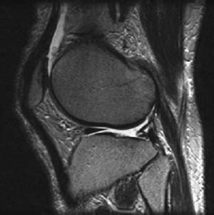
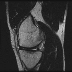
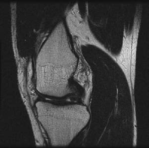
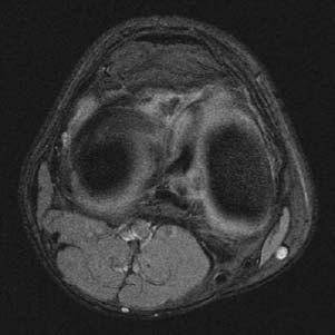
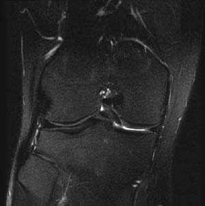
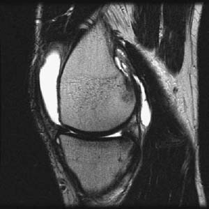
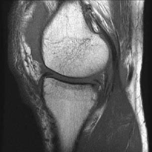
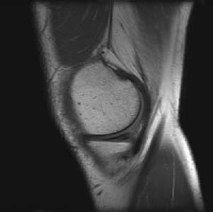
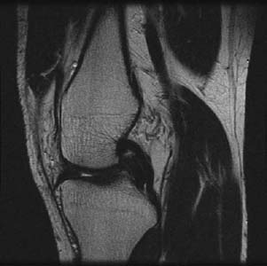
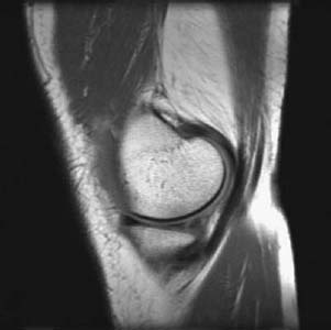
CASE 3 Hema N. Choudur, Anthony G. Ryan, and Peter L. Munk Five different patients presented with joint line pain. A history of trauma to the knee accompanied by an audible “pop” was given by the three younger patients. Limitation in knee flexion with no history of trauma was given by the two older patients. Figure 3A Figure 3B Figure 3C Figure 3D Figure 3F Figure 3G Figure 3H Figure 3I Figure 3J A T1-weighted sagittal image (Fig. 3A) shows a complex tear in the posterior horn of the medial meniscus. A T2-weighted sagittal image shows a complex tear in the posterior horn of the lateral meniscus (Fig. 3B). Figures 3C–3G show a flipped fragment of the posterior horn of the medial meniscus with the characteristic double posterior cruciate ligament sign. Sagittal T2-weighted (Fig. 3H) and sagittal T1-weighted (Fig. 3I) images show a missing segment of the posterior horn of the medial meniscus. A sagittal T1-weighted image (Fig. 3J) shows a linear oblique tear surfacing inferiorly in the posterior horn of the medial meniscus. Another sagittal T1-weighted image (Fig. 3K) shows a linear vertical tear in the posterior horn of the medial meniscus. Meniscal tears. None. The menisci are semilunar cartilages that serve to increase the congruency between the femoral condylar articular surfaces and the tibial plateau. The menisci serve various functions, including load transmission, stability, joint proprioception, shock absorption, and articular cartilage nutrition. The medial meniscus is thicker than the lateral, with the anterior horn about one third the thickness of the posterior, covering ~50% of the tibial plateau. The lateral meniscus is of uniform thickness anteroposteriorly and covers ~75% of the tibial plateau. When viewed from above, the meniscus has a crescent shape (meniscus means “little moon” in Greek). The meniscal ring is thickest at the periphery and tapers off centrally, creating a shallow cup to hold the rounded condyles of the femur. In cross section, the meniscus has a triangular wedge shape.
Meniscal Tear
Clinical Presentation

Radiologic Findings
Diagnosis
Differential Diagnosis
Discussion
Background
Related posts:
Stay updated, free articles. Join our Telegram channel

Full access? Get Clinical Tree


