CASE 35 Brian Edward Reeves, Anthony G. Ryan, Peter L. Munk, and Thomas Pope A 30-year-old man presented with a history of flushing, nausea, and emesis. Physical exam revealed urticaria pigmentosa. Figure 35A Figure 35B Figure 35C An anteroposterior (AP) radiograph of the abdomen from a small bowel follow-through (SBFT) study (Fig. 35A) shows small mucosal nodules and diffuse thickening of the valvulae conniventes throughout the proximal small bowel. The AP lumbar spine radiograph (Fig. 35B) shows diffuse osteosclerosis throughout the lumbar spine. A radiograph of the hands (Fig. 35C) shows diffuse osteosclerosis throughout. Mastocytosis. Diffuse osteopenia may be seen in osteoporosis, osteomalacia, hyperparathyroidism, and plasma cell myeloma. Multiple osseous lytic lesions may simulate the appearance of osteoporosis, sickle cell anemia, and Gaucher’s disease. Diffuse osteosclerosis may be observed in fluorosis, renal osteodystrophy, osteopetrosis, myelofibrosis, metastatic disease, and Paget’s disease. Mastocytosis comprises a group of disorders caused by an overabundance of mast cells, which are located predominantly in the skin, the linings of the stomach and small intestine, and the lungs. Mast cells release histamine and play important roles in wound healing, in blood vessel growth, and in the immune defense system. There are two general types of mastocytosis: cutaneous and systemic. Cutaneous is the more common form and mostly affects children. Mastocytosis occurs in children 75% of the time, is usually mild, and is often self-limited.
Mastocytosis
Clinical Presentation
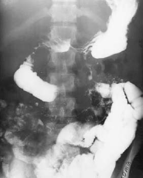
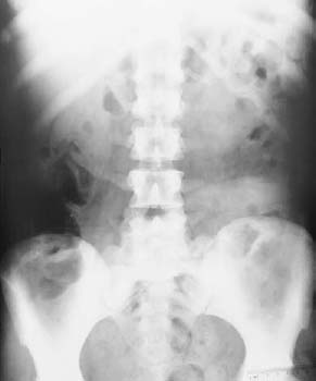
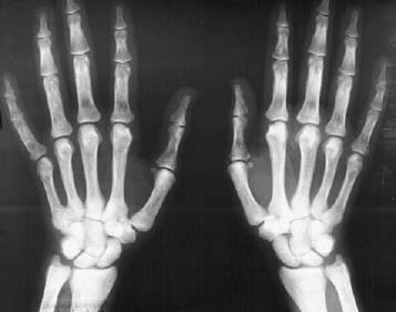
Radiologic Findings
Diagnosis
Differential Diagnosis
Discussion
Background
Related posts:
Stay updated, free articles. Join our Telegram channel

Full access? Get Clinical Tree


