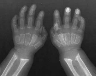
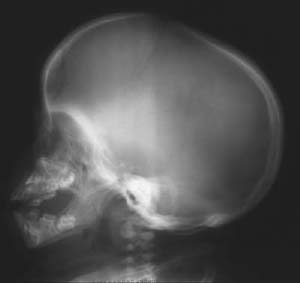
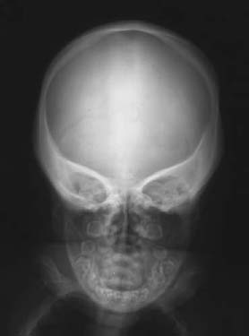
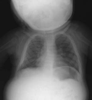
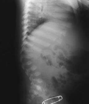
CASE 36 Brian Edward Reeves, Anthony G. Ryan, Peter L. Munk, and Thomas Pope A 1-year-old child developed coarse, thick, facial features, prominent dark eyebrows, cloudy corneas, progressive stiffness, gibbus deformity, and obvious mental retardation. The child had appeared normal at birth. Laboratory values showed increased urinary excretion of dermatan and heparan sulfates. Figure 36A Figure 36B Figure 36C Figure 36D Figure 36E Figure 36A shows pointing of the proximal portions of the second through fifth metacarpals, widening of the proximal and middle phalanges, and a V-shaped deformity of the distal radius and ulna. Figures 36B and 36C show macrocephaly, poorly developed mastoids, a prominent forehead, heavy supraorbital ridges, an elongated and J-shaped pituitary fossa, poorly developed paranasal sinuses, a large tongue, and a thick diploic space. Figure 36D shows widening of the anterior aspect of the ribs. Focal kyphosis at L2 due to the presence of a hypoplastic, bullet-shaped vertebral body is seen in Fig. 36E. Hurler’s syndrome.
Hurler’s Syndrome
Clinical Presentation
Radiologic Findings
Hands
Skull
Chest
Spine
Diagnosis
Discussion
Background
Related posts:
Stay updated, free articles. Join our Telegram channel

Full access? Get Clinical Tree


