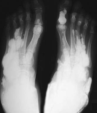
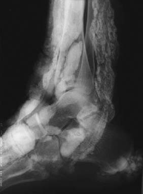
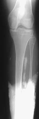
CASE 39 Anthony G. Ryan and Peter L. Munk A 65-year-old woman presented to her family practitioner with a recurrent history of progressive weakness, having particular difficulty climbing stairs. On examination, the patient had a striking violaceous discoloration and puffiness around her eyes and erythematous raised plaques on the skin overlying her knuckles. The practitioner noticed that the patient labored over standing up from the chair to get on the examination couch. Figures 39A Figures 39B Figure 39C Conventional radiographs of the feet (Fig. 39A), right ankle (Fig. 39B), and distal foreleg (Fig. 39C) demonstrate sheets and conglomerations of calcification within the subcutaneous soft tissues, some of which assume the configuration of underlying tendons and musculature. Dermatomyositis. Soft-tissue calcifications The differential for sheets of calcification within the soft tissues is essentially limited to two entities: Thought of as an idiopathic autoimmune condition affecting the skin and skeletal muscle, dermatomyositis may also affect cardiac muscle, joints, and lungs. It is a rare condition, with only five new cases per million expected per year. It has a bimodal age distribution affecting children (5–14 years) and older middle-aged adults (45–65 years), women twice as often as men. In the childhood group, subcutaneous calcifications and necrotizing vasculitis tend to predominate, whereas in the older age group, there is a well-established association with solid tumors (especially of the stomach, colon, and lung), the risk being 4.4 times that of an age-controlled group. This approximates to 1 in 10 patients with dermatomyositis having an underlying malignancy. The diagnosis of dermatomyositis in a person over 40 should prompt a search for an occult malignancy. Surveillance is also suggested, as the malignancies may take up to 5 years to declare themselves. Dermatomyositis rarely may be associated with other connective tissue disorders, for example, mixed connective tissue disorder and scleroderma. This is an autoimmune cell-mediated myopathy. The immune component is postulated to have an infectious origin, with protozoa (Toxoplasma gondii) and viruses (Coxsackie B RNA has been isolated from muscle biopsy specimens in one study in as many as 50% of dermatomyositis patients) under investigation. Cytotoxic T cells dominate the cellular inflammatory response; however, a humoral contribution is also evident. Microvasculature changes include deposits of immune complexes in vessel walls and the presence of microtubuloreticular structures in endothelial cells consistent with an accompanying vasculitis. The classic presentation is with a symmetric proximal myopathy as evinced by difficulty arising from the seated position and in reaching above the head.
Dermatomyositis
Clinical Presentation


Radiologic Findings
Diagnosis
Differential Diagnosis
METASTATIC
CALCINOSIS (NORMAL CALCIUM)
DYSTROPHIC
Discussion
Background
Etiology
Pathophysiology
Clinical Findings
Stay updated, free articles. Join our Telegram channel

Full access? Get Clinical Tree


