4 Arthroscopy
Arthroscopy is an invasive examination technique providing a direct view into the radiocarpal, the midcarpal, and the distal radioulnar joint. With arthroscopy, the condition of the articular cartilage, the ligaments, and the synovial structures can be assessed. In addition to evaluation of joint abnormalities, therapeutic interventions can be carried out in the same arthroscopy session.
Whereas arthroscopy quickly became established for the large joints, endoscopy of the small joints was first made possible by the development of smaller instruments that opened up new diagnostic and therapeutic perspectives in arthroscopy. Better knowledge of the complex anatomy of the wrist and increasing experience in the technical performance of arthroscopy helped to move arthroscopy out of the experimental stage into common use in hand surgery. Today it is indispensable in the treatment of wrist problems.
Necessary Equipment
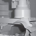
Because very little space is available for the arthroscope in those parts of the joint that are of arthroscopic interest, it is necessary to distend the joint cavity. For this purpose, with the arm bent at a right angle, a fixation device is attached to the hand, just as in the repositioning of a distal radius fracture ( Fig. 4.1 ). Fastening a weight to the upper arm creates tension in the opposite direction, which widens the joint space. Alternatively, a traction tower can be used, in which the arm is placed at the proximal and distal ends and stretched and can easily be moved for open interventions. The horizontal extension permits a quick exchange between arthroscopic and surgical procedures and is especially suited for arthroscopically controlled repositioning of intra-articular radius fractures.
For arthroscopy of the wrist, lenses with a shank diameter between 1.7 and 2.7 mm and with an inclination of 30° at the tip of the instrument are preferable. Introduction is achieved by means of a hollow cannula with a maximal outside diameter of 3.5 mm.
There is an intermediate space between the inner and the outer shank to allow fluid or gas to be introduced while the lens is in place. For purely diagnostic arthroscopy, the use of carbon dioxide as a contrast medium has the advantage of a more intensive contrast and correspondingly higher-quality images. For therapeutic interventions, however, fluid must be used to flush the joint.
The arthroscope is attached to video equipment, which consists of a mounted camera, a control unit, a light source, and a monitor.
Examining hooks (“probes”) of different lengths are also necessary for diagnostic purposes. For therapeutic interventions, special biopsy and gripping forceps, as well as motorized shaver systems with different heads, are used. Results are preferably recorded digitally. Connecting a personal computer with an integrated video card makes it possible to take digital freeze-frames and video sequences directly via the camera controls. Archiving is performed on CD-ROM or over a network card in the electronic patient record. Unlike a video printout, the arthroscopic images are always available and are of higher quality.

Arthroscopic Access
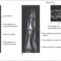
When planning arthroscopic access, the exact anatomical conditions of the wrist with its numerous tendons, nerves, and blood vessels must be taken into consideration. All accesses to the radiocarpal joint are on the dorsal side between the individual compartments of the extensor tendons. The accesses are named according to their continuous numbers. The distal radioulnar joint is only rarely examined arthroscopically. The standard accesses are listed in Table 4.1 and Figure 4.2
The 3–4 port is most often used to introduce the diagnostic lens, while the 4–5 port or the 6-R port serves as access for therapeutic instruments such as forceps or the shaver.
A superficial slit incision is made over the planned access point, which is then bluntly widened into a working canal. The next step is puncture with a hollow cannula and trocar, which is removed after correct placement and replaced with the arthroscope. The joint is filled with fluid or carbon dioxide through the introducer lock.

Related posts:
Stay updated, free articles. Join our Telegram channel

Full access? Get Clinical Tree


