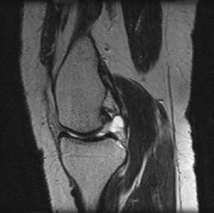
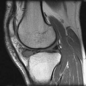
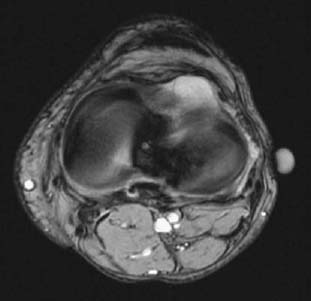
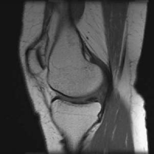
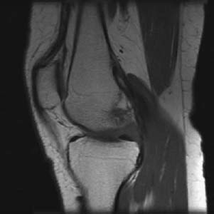
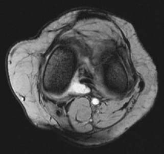
CASE 4 Hema N. Choudur, Anthony G. Ryan, and Peter L. Munk Two patients presented with focal knee pain, anterolateral in the first and posteromedial in the second, with a palpable mass in the latter. Figure 4A Figure 4B Figure 4C Figure 4D Figure 4F T1-weighted sagittal (Fig. 4A) and T2-weighted axial (Fig. 4B) images reveal a meniscal cyst arising from the anterior horn of the lateral meniscus. T1-weighted sagittal (Figs. 4C, 4D), T2-weighted sagittal (Fig. 4E), and T2-weighted axial (Fig. 4F) images reveal a parameniscal cyst arising from the posterior horn of the medial meniscus. In the first case, the T2-weighted images show a well-defined cystic lesion within the anterior horn of the lateral meniscus. A similar lesion was noted extending from the posterior horn of the medial meniscus in the second. An associated meniscal tear was evident in both. Meniscal cyst. The clue to the diagnosis lies in demonstrating the fluid nature of the cyst and the associated horizontal tear in the meniscus with which it communicates. Meniscal cysts were first described by Ebner in 1904. In 1923, the first series of meniscal cysts were reported as seen in 1% of meniscectomies. Magnetic resonance imaging (MRI) has revolutionized the detection and imaging of meniscal cysts. Meniscal cysts occur twice as often in men as in women—at an average age of 30 years. Lateral meniscal cysts are more common than medial by a ratio of 3:1.
Meniscal Cyst
Clinical Presentation
Radiologic Findings
Diagnosis
Differential Diagnosis
Discussion
Background
Stay updated, free articles. Join our Telegram channel

Full access? Get Clinical Tree


