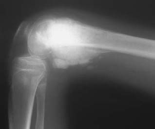
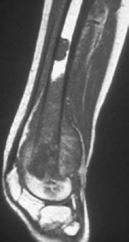
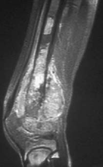
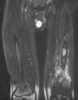
PART III Tumors George Nomikos, Anthony G. Ryan, Peter L. Munk, Mark Murphey A 14-year-old boy presented with an enlarging, painful, left distal femoral mass. Figure 41A Figure 41B Figure 41C Figure 41D The radiograph shows a large destructive mass arising from the distal femoral metaphysis (Fig. 41A). Dense matrix mineralization is apparent in both the intraosseous and soft-tissue components of the lesion. The pattern of mineralization is cloudlike, compatible with osteoid mineralization. Also note the aggressive-appearing periosteal reaction and Codman’s triangle. Sagittal T1-weighted images before (Fig. 41B) and after (Fig. 41C) intravenous contrast administration and a T2-weighted image (Fig. 41D) show a large area of marrow replacement in the distal femur and the associated soft-tissue component. In addition, there is a second, smaller focus of tumor in the femoral metaphysis separated from the primary tumor by normal intervening marrow. High-grade intramedullary (conventional) osteosarcoma with a skip metastasis. None. Osteosarcoma is the second most common malignant primary osseous neoplasm following myeloma. Although there are multiple subtypes of osteosarcoma, the conventional (or central intramedullary) type depicted in this case is the most common form. The most common skeletal location of the conventional osteosarcoma is about the knee, accounting for 50 to 55% of cases. Most affected patients are in their teens or twenties and there is a 2:1 male-to-female ratio. Lesions are most commonly metaphyseal, although diaphyseal (2 to 11%) and epiphyseal (< 1%) lesions may occur. Epiphyseal extension of metaphyseal lesions is common. Pathologically, these are malignant tumors of mesenchymal origin whose cells produce osteoid or immature bone. Some lesions contain areas of chondroid (5 to 25%) and fibroblastic-fibrohistiocytic differentiation (7 to 25%); however, most lesions are predominantly osteoblastic (50 to 80%).
CASE 41
Osteosarcoma
Clinical Presentation
Radiologic Findings
Diagnosis
Differential Diagnosis
Discussion
Background
Pathology
Imaging Findings
RADIOGRAPHY
COMPUTED TOMOGRAPHY
Related posts:
Stay updated, free articles. Join our Telegram channel

Full access? Get Clinical Tree


