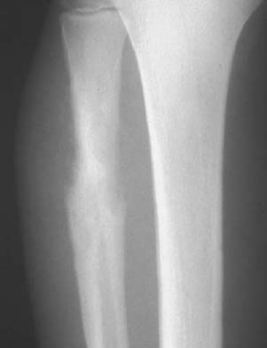
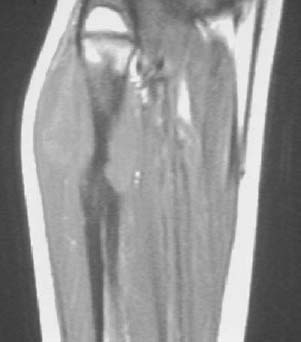
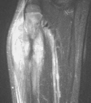
CASE 43 George Nomikos, Anthony G. Ryan, Peter L. Munk, and Mark Murphey A skeletally immature patient presented with a rapidly enlarging calf mass. Figure 43A Figure 43B Figure 43C An anteroposterior radiograph (Fig. 43A) of the proximal lower leg shows a poorly marginated area of permeative bone destruction as well as mild sclerosis involving the proximal fibular diaphysis. Note the marked cortical destruction. No mineralized matrix is identified. Also note the aggressive periosteal reaction along the diaphysis adjacent to the area of cortical destruction. A T1-weighted MRI (Fig. 43B) shows a large destructive mass arising from the proximal fibular diaphysis with formation of a large soft-tissue component. There is marrow replacement in the fibular shaft extending into the metaphysis. There is no evidence of extension across the physis. There is diffuse and heterogeneous enhancement of the mass after intravenous contrast administration (Fig. 43C). Ewing’s sarcoma.
Ewing’s Sarcoma
Clinical Presentation
Radiologic Findings
Diagnosis
Differential Diagnosis
Discussion
Background
Related posts:
Stay updated, free articles. Join our Telegram channel

Full access? Get Clinical Tree


