43 Magnetic Susceptibility–Related Artifacts on MRI
43.1 General Magnetic Susceptibility–Related Artifacts
43.1.1 Case 1 Presentation
A patient with a history of ventriculoperitoneal shunt presents with a headache. After a lateral skull film was obtained to evaluate the shunt valve and type, a brain MRI was performed.
43.1.2 Imaging Findings
The lateral view of the skull reveals a Strata NSC valve (▶ Fig. 43.1).

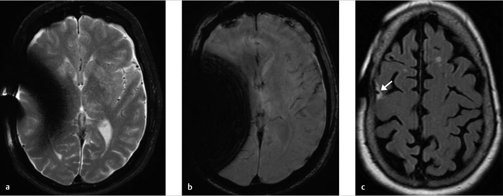
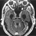
The MR images (▶ Fig. 43.2) reveal marked loss of signal on these T2-weighted images (T2WIs) due to substantial dephasing of signal that results from magnetic susceptibility artifact generated by the metallic portion of the valve. The artifact is substantially worse on susceptibility weighted imaging (SWI; ▶ Fig. 43.2b) than it is on the fast spin echo (FSE) T2WIs (▶ Fig. 43.2a) due to multiple rephasing 180-degree radiofrequency (RF) pulses in the latter sequence, which refocuses spins that have been dephased. The fluid-attenuated inversion recovery (FLAIR) sequence images (▶ Fig. 43.2c) are particularly vulnerable to local field inhomogeneity as the 180-degree inversion pulse is slice and therefore frequency specific. Artifactual loss of cerebrospinal fluid (CSF) suppression adjacent to the posterior aspect of the right middle frontal gyrus (arrow in ▶ Fig. 43.2c) is seen some distance cranially from the shunt valve as the presence of the metallic valve exerts its influence on magnetic field homogeneity far beyond the images on which it is present.
Cause: Magnetic susceptibility artifact due to metallic shunt components.
43.1.3 Essential Facts about Magnetic Susceptibility Artifacts in MR Imaging (www.MRIquestions.com)
The signal intensity of any given tissue depends upon many factors including proton density, T1 and T2 of the tissue, echo time (TE) and TR (repetition time) of the imaging sequence, and local magnetic field homogeneity.
The signal intensity of tissues is markedly influenced by local magnetic field inhomogeneity especially on T2WIs that are of the gradient recalled echo (GRE) type of sequence as opposed to spin echo–type sequences.
GRE, MPGR, SPGR, most echo planar, diffusion-weighted images (DWIs), and SWI are sequences that are particularly susceptible to loss of signal when there is an inhomogeneous magnetic field.
Substances with unpaired electrons in their outer shell (deoxyhemoglobin, methemoglobin, ferritin, hemosiderin, Fe, Ni, Co) locally augment the magnetic field strength, which leads to field inhomogeneity and therefore a difference in frequency of the spin of protons. This leads not only to loss of phase, which means there will be a significant decrease in signal intensity on T2WIs, but also to incorrect spatial localization of signal. Misregistration of detected signal whose frequency has been altered by magnetic susceptibility artifact typically results in bright lines from signal “pile-up” and dark lines, due to signal being displaced.
Bone–air or soft tissue/bone–air interfaces also distort the local magnetic field and result in similar artifacts.
Although metal artifact may not be completely eliminated on MRI, the following techniques may reduce its effect:
View-angle tilting.
Using thinner slices.
Using higher bandwidth imaging.
Lower field strength magnet.
Using short tau inversion recovery (STIR) rather than chemical fat saturation techniques.
Use of FSE technique and avoidance of GRE-type imaging.
43.1.4 Companion Cases
Companion Case 1: The Use of Metal Suppression Techniques in MRI to Reduce Metal Artifacts
History
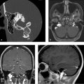
Brain MRI was obtained for the evaluation of headaches in a patient with a metallic pellet present in the ethmoid sinus.
Imaging Findings
Sagittal T1WI (▶ Fig. 43.3a) reveals substantial dephasing artifact that obliterates visualization of the paranasal sinuses even on the FSE-T2WIs (▶ Fig. 43.3b,c).

Sagittal T1WI (▶ Fig. 43.4 a) from a different patient’s brain MRI performed on the same magnet shows substantially worse artifact than that present in the first patient’s images (▶ Fig. 43.3) due to a nonremovable dental appliance. However, use of metal suppression techniques dramatically reduces the metallic associated artifact on the repeat sagittal T1WI (▶ Fig. 43.4b) and also on the axial FSE-T2WI (▶ Fig. 43.4c).

Companion Case 2: Pigmented Cosmetics May Result in MRI Artifacts
History
Young woman’s brain MRI plus special views of the orbits was ordered for a history of headache and blurred vision.
Imaging Findings
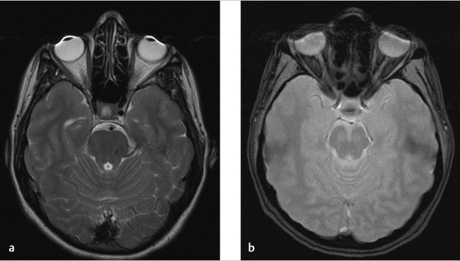
The MRI technologist noted magnetic susceptibility artifact present in association with the patient’s eyelids on the brain MRI (▶ Fig. 43.5) prior to the orbit-specific images. The patient confirmed that, as she had indicated on the MRI screening form, she did not have either tattooed eyeliner or gold weights implanted for facial nerve injury. She was, however, wearing heavy purple eyeliner that she subsequently removed, and the scan was repeated showing elimination of this artifact (▶ Fig. 43.6a,b).

Companion Case 3: Large, Extensively Pneumatized Sinuses and Petrous Apices May Increase Bone–Soft Tissue/Air Interface Related Susceptibility Artifact
History
Brain MRI was obtained for possible microvascular ischemic disease in an older male patient.
Related posts:
Stay updated, free articles. Join our Telegram channel

Full access? Get Clinical Tree


