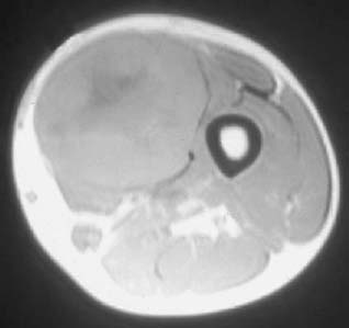
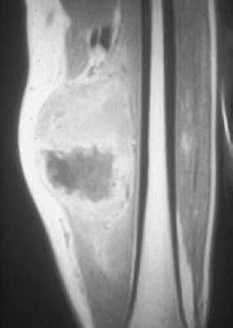
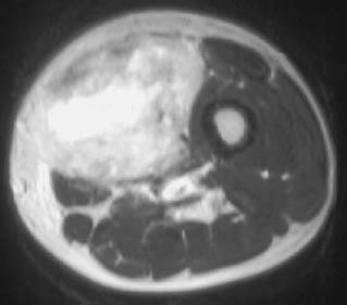
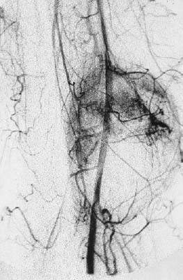
CASE 44 George Nomikos, Anthony G. Ryan, Peter L. Munk, and Mark Murphey A 40-year-old man presented with an 8-month history of a painless thigh mass. Figure 44A Figure 44B Figure 44C Figure 44D Axial T1- (Fig. 44A), postcontrast coronal T1- (Fig. 44B), and axial T2- (Fig. 44C) weighted images show a large intramuscular mass arising in the vastus medialis muscle. The lesion has a nonenhancing central area that is low signal on the T1-weighted image and high signal on the T2-weighted image, compatible with a region of central necrosis. The peripheral solid portion of the lesion is moderately heterogeneous on the T2-weighted image and shows diffuse enhancement. No fat is seen in the mass. No mineralization was identified in the mass on CT (not shown). The angiogram (Fig. 44D) shows marked tumor blush and neovascularity in the arterial phase of perfusion. There is also slight narrowing of the femoral artery, compatible with tumor encasement. Primary soft-tissue malignant fibrous histiocytoma (MFH).
Malignant Fibrous Histiocytoma
Clinical Presentation
Radiologic Findings
Diagnosis
Differential Diagnosis
Discussion
Background
Related posts:
Stay updated, free articles. Join our Telegram channel

Full access? Get Clinical Tree


